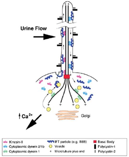
FIGURE 1.
Structure of primary cilia. The cilium protrudes from the apical surface of the cell. The axoneme, or cilium core, grows out from the basal body and is covered by ciliary membrane. The axoneme is composed of nine doublet microtubules nucleated directly from the basal body. Intraflagellar transport (IFT) particles transport proteins to the distal end of the cilium. Kinesin-II is the microtubule motor responsible for anterograde IFT. Retrograde transport utilizes dynein. Polycystin-2 is a calcium channel located on the primary cilium, where it interacts with polycystin-1.

