FIGURE 4.
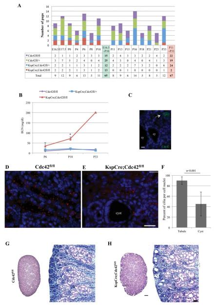
Lack of Cdc42 in kidney tubule cells leads to an early postnatal death due to lack of cilia and PKD. KspCre;Cdc42fl/+ mice (KspCadherin is expressed in distal tubules and collecting ducts) were mated to Cdc42fl/fl mice, and all four possible genotypes were readily identified by PCR (data not shown here). (A) Of 67 pups tested from P11-P33, there were 22 Cdc42fl/fl, 19 Cdc42fl/+, 24 KspCre;Cdc42fl/+ mice, with one dead (mostly eaten) homozygous Cdc42 knockout (KspCre;Cdc42fl/fl) mouse found at P15, and one long-surviving homozygous Cdc42 knockout (KspCre;Cdc42fl/fl) mouse that was euthanized at P33 due to failure to thrive, growth restriction, and renal failure. (B) By linear regression modeling, BUN levels in Cdc42 kidney-specific knockout mice were significantly higher than in littermate control KspCre;Cdc42fl/+ and Cdc42fl/fl mice at P10 (p = 0.035). (C) By immunofluorescence, Cdc42 is not seen in the cysts (cy), but is seen in the normal-appearing tubules of Cdc42 kidney-specific knockout mice (arrow). Bar = 10 μm. (D–F) Immunostaining of acetylated alpha tubulin (red) in kidneys from control (D) and Cdc42 kidney-specific knockout mice (E) at P4, shows lack of cilia in cells surrounding cysts. (F) Quantification from kidney tubule cell-specific Cdc42 knockout mice shows a significant decrease in the number of cilia seen per cell in cysts (which presumably are deficient in Cdc42, as no cysts were seen in control mice) compared to normal-appearing tubules, which most likely express Cdc42. Bar = 20 μm. (G, H) Hematoxylin/eosin stained sections of kidneys from control (G) and Cdc42 kidney-specific knockout mice (H) at P4 (left) and P6 (right). Bar for kidneys on left in G, H = 200 μm. Bar for kidney sections on right in G, H = 20 μm. (Reproduced with permission from the Journal of American Society of Nephrology).

