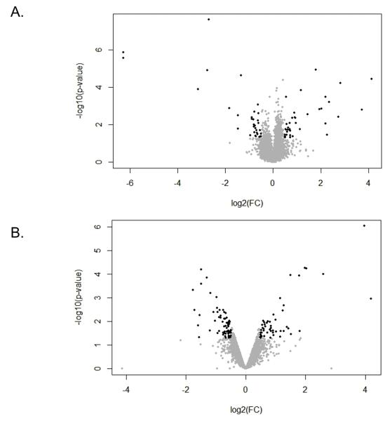
Figure 1. Volcano plot of genes differentially expressed in CD4+ T cells between BDC-Idd9.905 and BDC mice.
Volcano plot showing the relative abundance of transcripts in ex vivo (A) and p79-stimulated CD4+ T cells from BDC-Idd9.905 mice compared to BDC mice. Log2 of fold change (FC) is presented on the x-axis and −log10 of p values is represented on the y-axis. Transcripts that passed the cutoff of p<0.05 and FC>1.4 (log2=0.5) were considered to be differentially expressed and are shown in black. Genes that are significantly up-regulated in BDC-Idd9.905 CD4+ T cells compared to BDC CD4+ T cells are on the right, while down-regulated genes are on the left of FC=0. Grey dots indicate genes, which did not show differential expression between the two strains.

