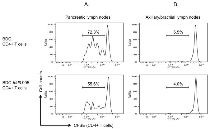
Figure 7. Impaired proliferation of BDC-Idd9.905 CD4+ T cells to endogenous autoantigen.
BDC-Idd9.905 and BDC CD4+ T cells were CFSE-labeled and transferred (5 × 10⁶, i.v.) into non-diabetic NOD mice. After 90 hours, proliferation of transferred CD4+ T cells from pancreatic lymph nodes (A) and from axillary/brachial lymph nodes (B) as control was determined by assessing CFSE dilution by flow cytometry. One of three independent experiments each with similar data is shown. Numbers in histograms represent percentages of CFSE+ CD4-gated T cells that underwent cell divisions.

