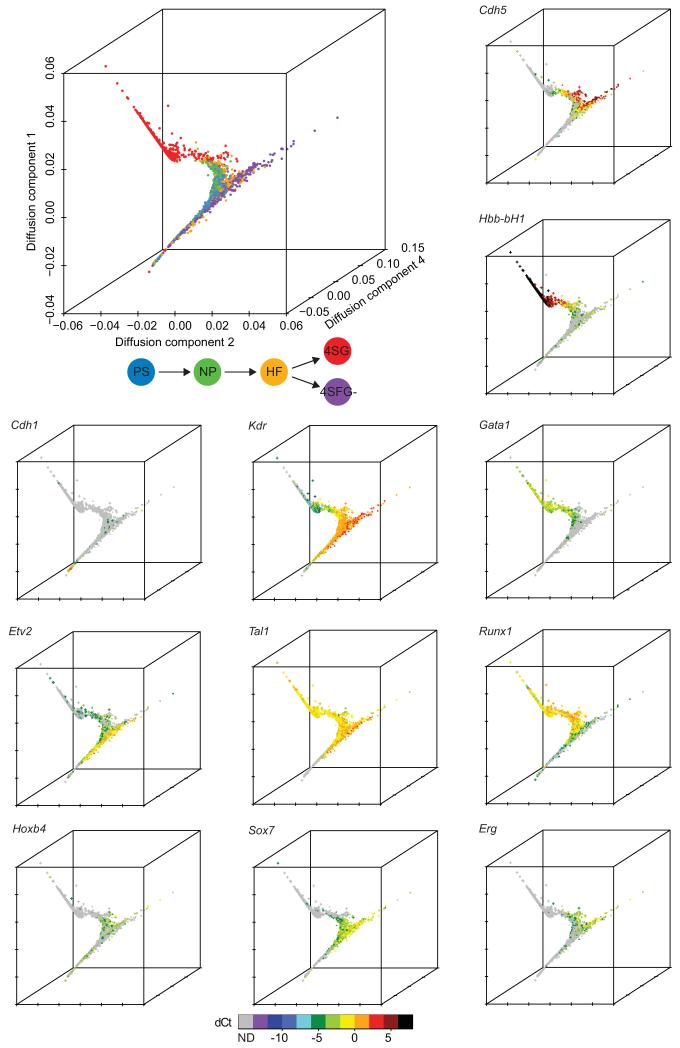
Figure 2. Diffusion plots identify developmental trajectories.
Diffusion plot of all 3934 cells calculated from the expression of 33 TFs and seven marker genes (top left). Blue, PS; green, NP; orange, HF; red, 4SG; purple, 4SFG−. The expression levels of individual genes were then overlaid onto the diffusion plot to highlight patterns of expression (see Supplementary Fig. 5 for additional genes). Circle, PS; diamond, NP; triangle, HF; cross, 4SG; square, 4SFG− (visible in high resolution version of figure).

