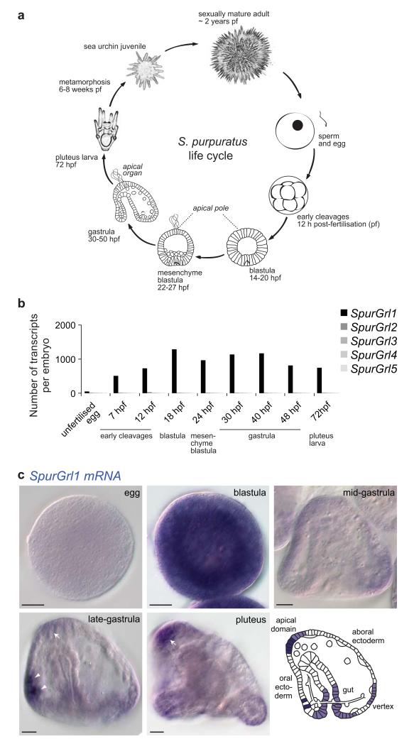
Figure 6. Early developmental expression of an S. purpuratus Grl.
(a) Schematic of the life cycle of the sea urchin S. purpuratus.
(b) Quantitative RT-PCR analysis of the temporal expression of the five SpurGrl genes during nine developmental time points. Data are represented as number of transcripts per embryo and represent the average of four technical replicates in each of two independent biological replicate samples (see Supplementary Table 3).
(c) RNA in situ hybridisation using a riboprobe against SpurGrl1 on whole mount S. purpuratus of five developmental stages. All the embryos represent lateral views: as indicated in the schematic, the oral ectoderm is on the left and the apical domain on the top in gastrula and pluteus specimens. The arrows mark the expression of SpurGrl1 in the apical domain (where the apical organ will form), while the arrowheads mark a pair of presumptive neurosecretory cells in the oral ectoderm. Scale bars = 20 μm.

