
Figure 2.
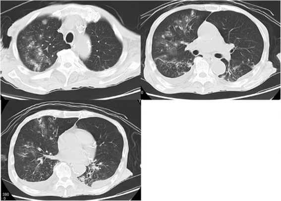
Multiple small cavitary lesions and nodules. Chest CT images obtained on the 14th day of admission showed multiple small cavitary lesions and nodules surrounded by ground-glass opacity, and also bilateral pleural effusion. These lung abnormalities seemed to be in a peribronchovascular distribution.