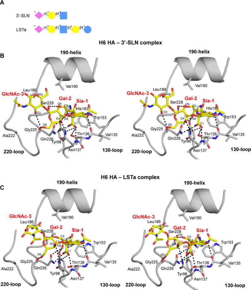
Figure 3. Crystal Structures of the H6 HA in Complex with Avian Receptor Analogs.

(A) Cartoon representation of the glycan structures of avian receptor analogs 3′-SLN and LSTa. Sia is the abbreviation for sialic acid, Gal for galactose, GlcNAc for N-acetylglucosamine and Glc for Glucose. (B and C) Stereo representations of the interactions between the HA H6 RBS and the avian receptor analogs. The conserved secondary elements of the HA RBS (130-loop, 190-helix and 220-loop) are labeled and shown in cartoon representation. Selected residues and receptor analogs are labeled and shown in sticks. The RBS is colored in gray and the receptor analogs in yellow. Hydrogen bond interactions of Sia-1 and Gal-2 and GlcNAc-3 of 3′-SLN (B) and LSTa (C) with the H6 HA RBS (see also Figures S3 and S4).
