
Figure 4. Crystal Structure of the H6 HA in Complex with a Human Receptor Analog.
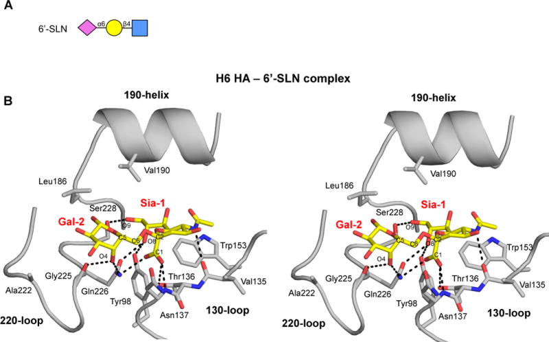
(A) Cartoon representation of the glycan structure of human receptor analog 6′-SLN. (B) Stereo representation of the hydrogen-bond interactions of Sia-1 and Gal-2 of 6′-SLN with the H6 HA RBS. The conserved secondary elements of the HA RBS (130-loop, 190-helix and 220-loop), as well as Tyr 98 and Trp153, are labeled and shown in cartoon representation (see also Figures S3 and S4).