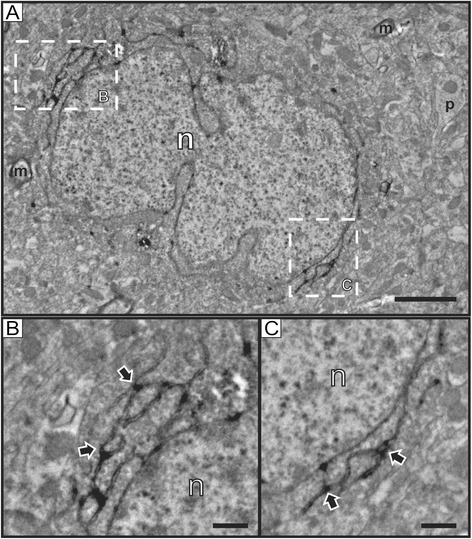
Figure 2.

Reelin neuronal labeling at the electron microscope. A) Low magnification electron microscopy image of a reelin immunolabeled neuron in the cortex. B-C) Detail images of the areas indicated with dashed-line boxes in A. Note that reelin labeling is specifically located in the rough endoplasmic reticulum (outlined black arrows). m: myelinated process; n: nucleus; p: neuronal process. Scale bars: 2 microns in A; 0.5 microns in B-C.
