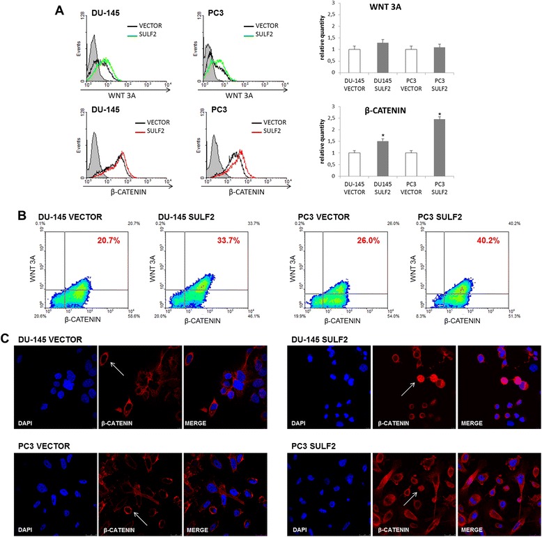
Figure 7.

WNT signaling pathway in SULF2 ovexpressing prostate cancer cells. DU-145 and PC3 cells were immunostained with WNT 3A and β-catenin antibodies (R&D) and analyzed by flow cytometry, as described in methods. The graphics represent relative quantity of positive cells (A). Representative pictures are shown, indicating the percent of double-stained cells (B). Prostate cancer cells were immunostained for β-catenin and analyzed by confocal microscopy. The nuclei (blue) were stained with DAPI. (C). (VECTOR: cells transfected with empty vector, SULF2: cells transfected with SULF2 expressing plasmid). *P ≤ 0.05.
