. 2015 Feb 24;4:e05118. doi: 10.7554/eLife.05118

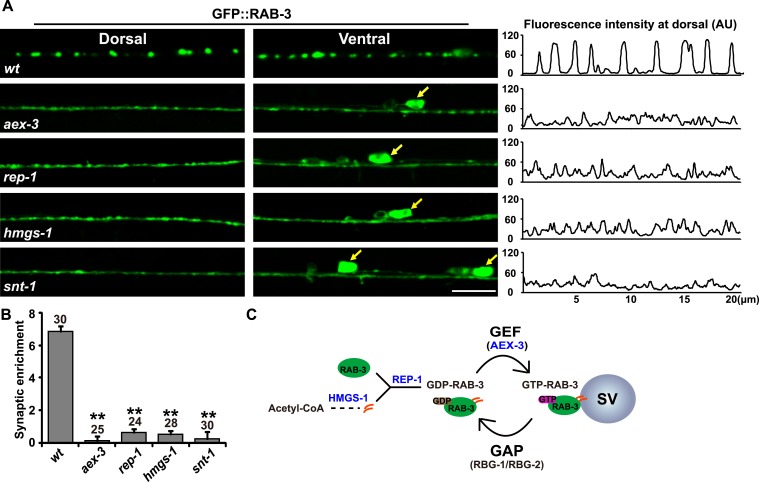
Figure 1. RAB-3 synaptic vesicle association requires SNT-1.

(A) Punctate distribution of GFP::RAB-3 in C. elegans motor neurons in wild-type animals (top). The GFP::RAB-3 puncta become diffuse in aex-3, rep-1, hmgs-1, and snt-1 mutants (lower panels). Yellow arrows indicate the cell bodies along the ventral cord. A representative line-scanning image for each genotype is shown in the right panel. (B) Quantification of the synaptic enrichment in wild-type, aex-3, rep-1, hmgs-1, and snt-1 animals. Data are presented as mean ± SD; **p < 0.01. (C) Schematic representation of the RAB-3/SV association and dissociation cycle. Scale bar, 5 µm.

DOI: http://dx.doi.org/10.7554/eLife.05118.003

Figure 1.

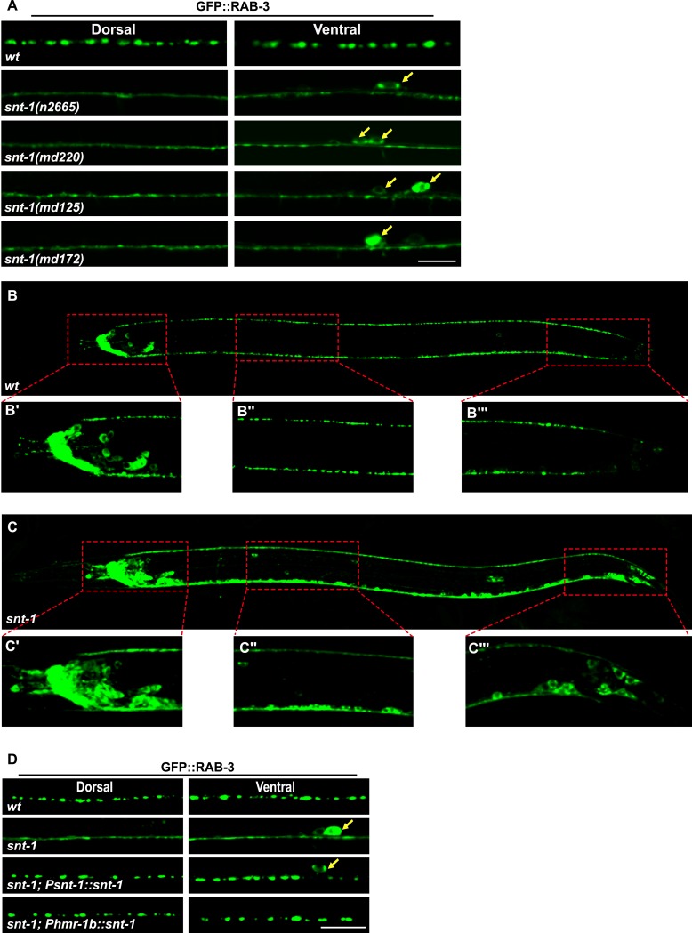
Figure 1—figure supplement 1. snt-1 is require for RAB-3 synaptic vesicle localization.

Figure 1—figure supplement 1.

(A) Multiple snt-1 alleles show the diffuse GFP::RAB-3 phenotype. White arrows indicate the cell bodies. (B) GFP::RAB-3 expressed pan-neuronally under the control of the Prab-3 promoter displays punctate distribution. (C) GFP::RAB-3 is diffuse in all Prab-3-expressing cells. Red boxed areas are enlarged in panels underneath the image taken from a whole animal (B′, B″, B‴, C′, C″ and C‴). (D) The diffuse GFP::RAB-3 phenotype in snt-1 mutants is rescued by expressing wild-type snt-1 gene pan-neuronally using the Psnt-1 promoter or in DD, VD, and AS neurons using the Phmr-1 promoter. Scale bar, 5 µm.