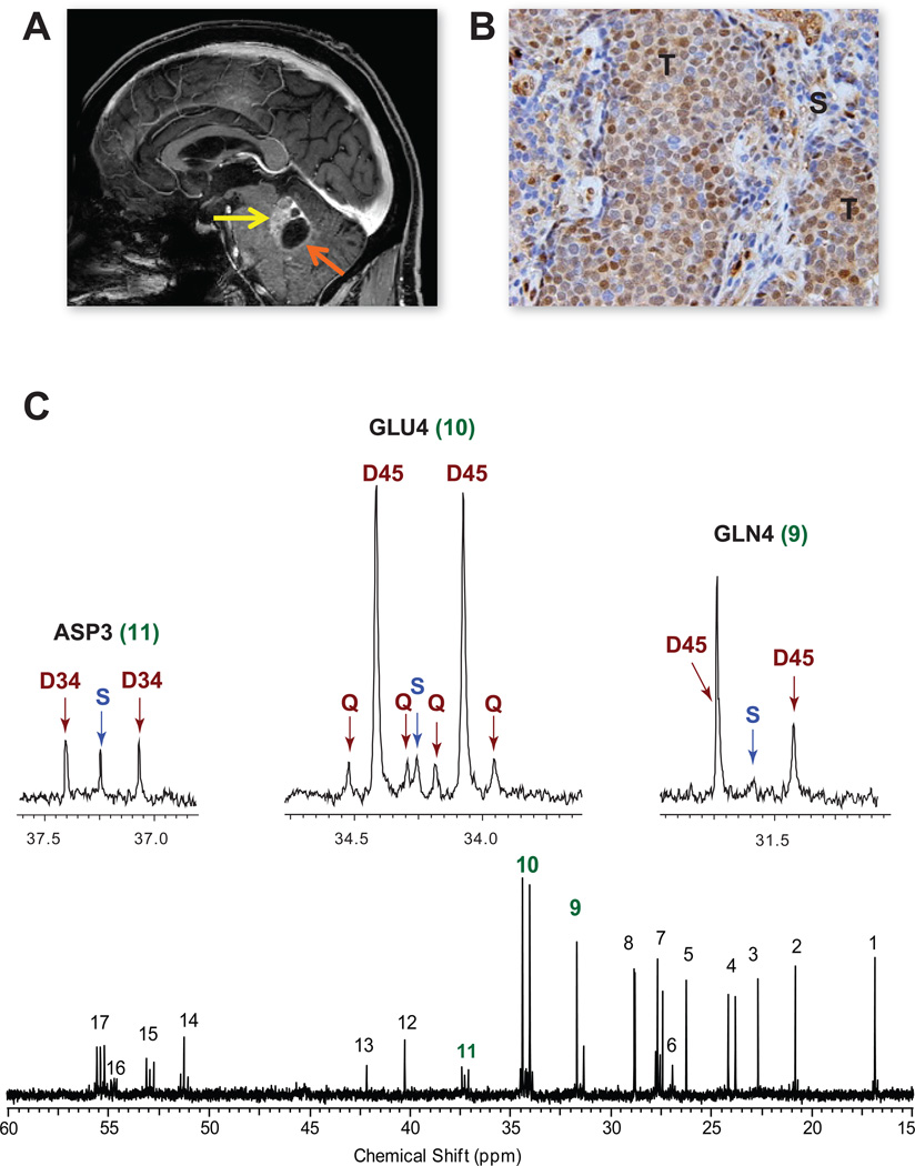
Figure 7. Infusion of [1,213C]acetate in a patient with a non-small cell lung cancer brain metastasis.
(A) Pre-operative MRI, sagittal image, shows a gadolinium-enhancing tumor in the left cerebellum (yellow arrow) with a cystic component (orange arrow). (B) Moderate ACSS2 immunoreactivity in the tumor (T) with lack of staining in the surrounding stroma (S). Scale bar 10 um. (C) 13C-NMR spectrum with GLU4, GLN4 and ASP3 insets. Abbreviations same as Figure 2. Chemical shift assignments same as in Figure 6 with the addition of 11, Aspartate C3; 13, Glycine C2; 14, Alanine C2; and 15, Aspartate C2. See also Figure S4 for 13C-spectra from 2 additional patient tumors.

