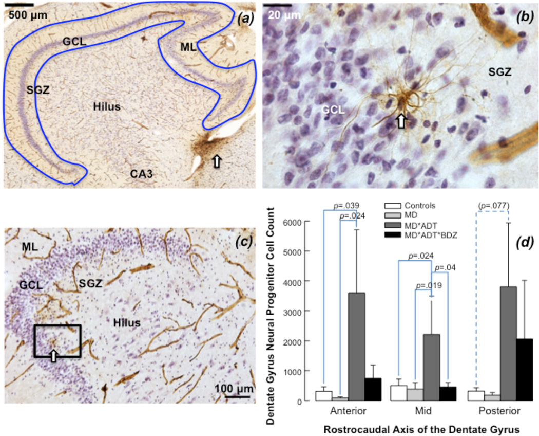
Figure 3. Neural progenitor cells in the human hippocampus.
(a) Nestin-immunoreactive neural progenitor cells (NPCs) and capillaries (in brown) were primarily found within the subgranular zone (SGZ) and in the subventricular zone (white arrow). Granule cells in the granule cell layer (GCL) and glia were stained for Nissl using Cresyl Violet. Capillaries are visible also in the hilus and Cornu Ammonis (CA) regions. The molecular layer (ML) is indicated. The blue outline defines the region of interest for cell counting with stereology. (b) NPCs are seen in the SGZ (white arrow). Capillaries are visible throughout the section. (c) Multipolar Nestin-immunoreactive NPC (enlargement of rectangle in b) shows dendrite processes extending into the GCL and touching the capillary located in the SGZ. Nestin-positive blood vessels display their characteristic appearance. (d) Nestin-immunoreactive NPC number in the dentate gyrus (DG). Antidepressant-treated subjects with mood disorders (MD*ADT) had more NPCs in the anterior and mid DG compared with untreated MD subjects (MD) and subjects with no psychopathology or treatment (Controls); subjects treated with antidepressants and benzodiazepines (MD*ADT*BZD) had fewer NPCs in mid DG compared with MD*ADT and similar number of NPCs as untreated MD in every DG subregion. Untreated MD did not show fewer NPCs than Controls in any DG subregion.

