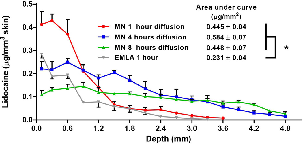
Fig. 7. Diffusion of lidocaine in porcine skin in vitro.
Solid dispersion coatings were formed on microneedles by dip coating in a molten solution containing polyethylene glycol (PEG) and lidocaine at 50% mass ratio. Six arrays of microneedles were used to deliver 81 µg lidocaine into porcine skin measuring 1 cm×1 cm, in vitro, by inserting microneedles at equal spacings and leaving them inserted for 3 min. Upon removal of microneedles, the skin tissues were incubated in a humid chamber for 1, 4 or 8h (n=3 skin tissues per time point). For comparison, 0.15 g EMLA cream was topically applied on skin for 1 h. Skin samples were subsequently sectioned to quantify lidocaine that diffuses along the depth of the tissue.

