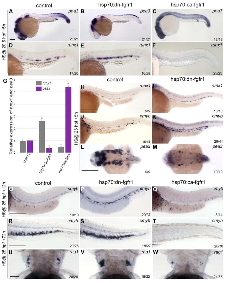
Figure 1. FGF signaling represses HSC formation and maintenance.
(A–F) Embryos were heatshocked at 20.5 hpf and analyzed at 26 hpf. (A–C) pea3 expression is downregulated in hsp:dn-fgfr1 (B) and upregulated in hsp70:ca-fgfr1 embryos (C) compared to controls (A). (D–F) Aortic expression of runx1 is enhanced in hsp70:dn-fgfr1 (E) and depleted in hsp70:ca-fgfr1 embryos (F) compared to controls (D). (G) Quantification of runx1 and pea3 mRNA expression in dissected trunks normalized to ef1α. Expression of each gene was set to 1 in the control. Data are shown as average ± s.d. values; p<0.001. (H–M) Embryos were heatshocked at 25 hpf and analyzed at 30 hpf. (H–I) runx1 expression is increased in hsp70:dn-fgfr1 embryos (I) compared with controls (H). Similar results are seen with cmyb expression in control (J) and hsp70:dn-fgfr1 embryos (K). (L and M) Dorsal view of pea3 expression in the head shows downregulation of pea3 upon depletion of FGF signaling. (O–W) Embryos were heatshocked at 25hpf and analyzed 12h and 72hpHS. (O–Q) cmyb expression is more intense in the hsp70:dn-fgfr1 embryos (P) compared to control embryos (O). Conversely, embryos in which FGF signaling is increased, display a drastic diminution of cmyb expression (Q). (R–S) Comparison of cmyb expression in the CHT of heatshocked transgenic embryos and controls. The augmentation of HSC numbers detected at 26hpf upon FGF modulation is maintained in the CHT of the hsp70:dn-fgfr1 embryos while the CHT of the hsp70:ca-fgfr1 embryos are devoid of cmyb cells. (U–W) Effect of FGF signal alteration on T-cells. FGF ablation (V) or augmentation (W) has opposite effects on rag1+ cells. Scale bars: (A) 50um, (D, H) 200um, (L, O, R) 250um, (U) 50um.

