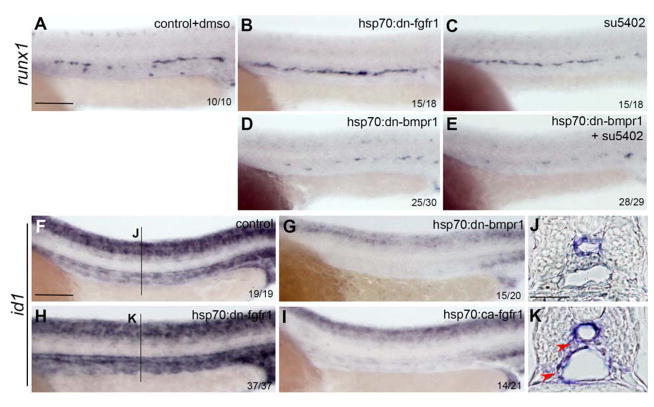
Figure 5. Epistatic analysis of BMP and FGF signaling interaction.
(A–C) runx1 expression is increased in hsp70:dn-fgfr1 embryos (B) and embryos treated with su5402 (C) compared to controls (A). Overexpression of hsp70:dn-bmpr1 impairs emergence of HSCs (D) compared to control (A) or FGF inhibited embryos (B and C). HSC emergence is not rescued in hsp70:dn-bmpr1 embryos following blockade of FGF signaling using su5402 (E). (F–P) id1 expression is reduced upon inhibition of BMP signaling (G) as well as augmentation of FGF signaling (K), compared to control embryos (F). In the absence of FGF signaling, id1 expression is increased in the vasculature and in some cells surrounding the vessels (N, and K, red arrows). Scale bars: (A, F) 100um, (J) 30um.

