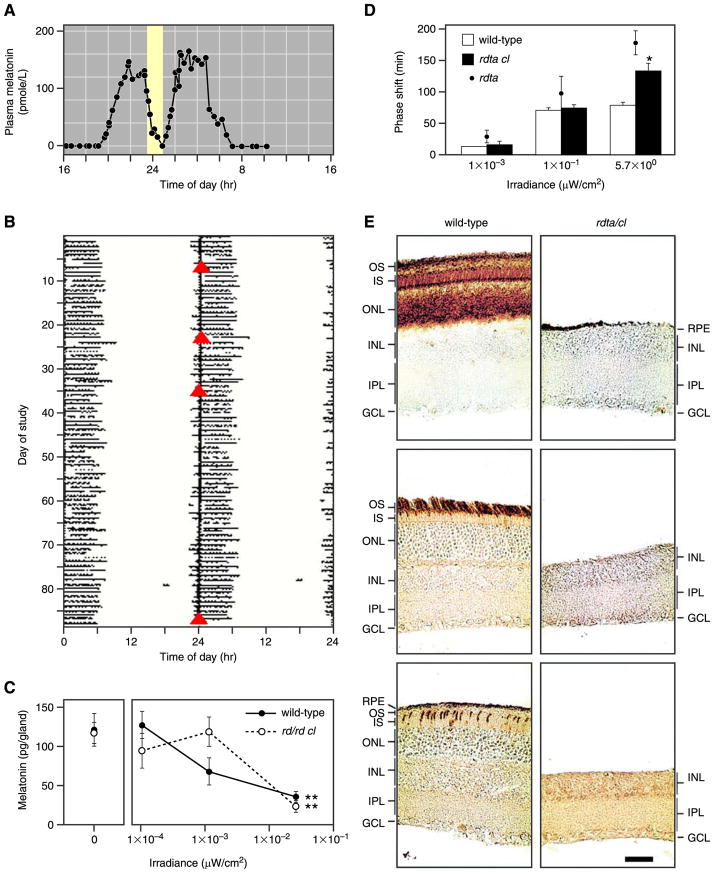
FIG. 1.
Photoreception without rods and cones. A: suppression of plasma melatonin by light (lighter region) in a patient who was blind from Leber’s congenital amaurosis and lacked a detectable electroretinogram. B: the sleep-wake pattern of the blind patient in A. Solid horizontal lines represent periods of sleep, and open triangles (red) indicate times of peak plasma melatonin level (a marker of the patient’s endogenous circadian period). This patient’s activity patterns were entrained to the environmental 24-h cycle. [A and B modified from Czeisler et al. (34).] C: suppression of melatonin synthesis in wild-type and rodless/coneless mice by light of different irradiances. [Modified from Lucas et al. (115).] D: shift of circadian phase in wild-type, rodless/coneless (rdta/cl), and rodless (rdta) mice by light of different irradiances. E: retinal cross-sections of wild-type and rodless/coneless (rdta/cl) mice stained with antibodies recognizing rod pigment (top), rod and green cone pigments (middle), and ultraviolet cone pigment (bottom) to demonstrate that no outer retinal photoreceptors are detectable in rodless/coneless mice. The same was observed for retinas of rd/rd cl mice used for C. OS, outer segments; IS, inner segments; ONL, outer nuclear layer; INL, inner nuclear layer; IPL, inner plexiform layer; GCL, ganglion cell layer; RPE, retinal pigment epithelium. Scale bar is 40 μm. [D and E modified from Freedman et al. (52).]

