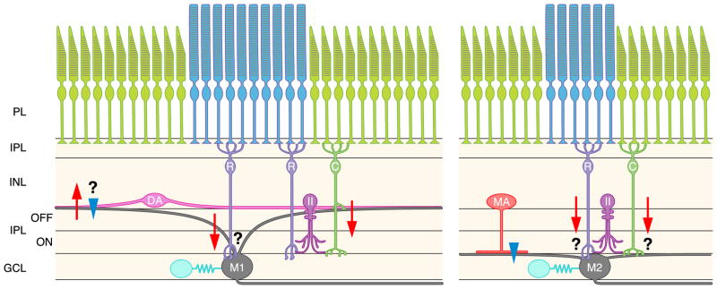
FIG. 10.
Schematic of ipRGCs and retinal circuitry. A schematic of the retinal cross-section, illustrating the reported circuitry of M1 (left) and M2 (right) ipRGCs. Downward-pointing arrows indicate transmission to ipRGCs, and upward-pointing arrows indicate transmission from ipRGCs. Red arrows indicate excitation, and blue arrowheads indicate inhibition. Question marks indicate connections/interactions awaiting confirmation from electrophysiology. M1, M1 ipRGC; M2, M2 ipRGC; R, rod bipolar cell; C, cone bipolar cell; II, AII amacrine cell; MA, monostratified amacrine cell; DA, dopaminergic amacrine cell; PL. photoreceptor layer; IPL, inner plexiform layer; INL, inner nuclear layer; GCL, ganglion cell layer. IpRGCs are also coupled electrically to cells in the GCL (blue cells). The retinal circuitry of M3 cells has not been reported and thus is not illustrated here.

