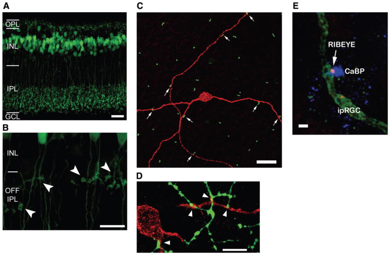
FIG. 11.
ON synapses in the OFF sublamina to ipRGCs. A: vertical section through a mouse retina expressing GFP under control of the mGluR6 promoter, active in ON cone bipolar cells. Scale bar is 20 μm. B: high-power view of section in A to show axonal swellings, which are en passant synapses, and lateral extensions from the axons terminating in swellings, which are ectopic synapses. Scale bar is 10 μm. [A and B modified from Dumitrescu et al. (43).] C: melanopsin immunostaining of a flat-mounted rabbit retina (red). Descending axons of calbindin-expressing bipolar cells (green) forming ectopic, en passant synapses on ipRGC dendrites in the OFF sublamina of the IPL. Scale bar is 20 μm. D: axon terminals of calbindin-expressing bipolar cells in rabbit (green) contacting ipRGC dendrites in the ON sublamina of the IPL. Scale bar is 10 μm. E: synaptic ribbons (positive for RIBEYE-immunostaining) colocalized with junctions between calbindin-positive axons (blue) and ipRGC dendrites (green). Scale bar is 1 μm. [C–E modified from Hoshi et al. (94).]

