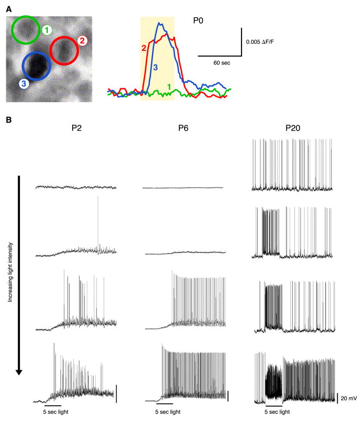
FIG. 13.
IpRGCs during development. A: calcium imaging of a retina taken from wild-type mouse at birth. Two of three cells studied in this field of view (left) showed a light-driven rise in calcium (right). Timing of the light stimulus shown by shaded region. [Modified from Sekaran et al. (178).] B: increase in photosensitivity of ipRGCs with age. Whole cell, current-clamp recordings from mouse ipRGCs in the flat-mount retina with synaptic transmission intact. Responses to four different light intensities are shown for three developmental times. [Modified from Schmidt et al. (174).]

