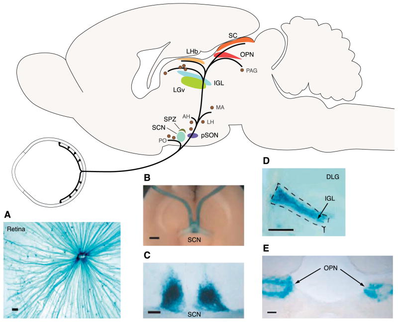
FIG. 3.
Brain targets of ipRGCs. A schematic of the mouse brain in sagittal view showing a sampling of regions innervated by ipRGCs. [Modified from Hattar et al. (87).] PO, preoptic area; SCN, suprachiasmatic nucleus; SPZ, subparaventricular zone; pSON, peri-supraoptic nucleus; AH, anterior hypothalamic nucleus; LH, lateral hypothalamus; MA, medial amygdaloid nucleus; LGv, ventral lateral geniculate nucleus; IGL, intergeniculate leaflet; BST, bed nucleus of the stria terminalis; LGd, dorsal lateral geniculate nucleus; LHb, lateral habenula; SC, superior colliculus; OPN, olivary pretectal nucleus; PAG, periaqueductal gray. A: flat-mount retina of a mouse with the tau-lacZ marker gene targeted into the melanopsin gene locus (opn4+/− tauLacZ+/−). Blue color shows X-gal staining of the β-galactosidase activity coded by tau-lacZ in the ipRGCs. IpRGC axons can be seen coursing to the optic disc. Scale bar is 100 μm. [Modified from Hattar et al. (89).] B: ventral view of the opn4+/− tauLacZ+/− brain showing ipRGC axons running in the optic nerve and innervating the suprachiasmatic nuclei (SCN). Scale bar is 1 mm. C: coronal section of the opn4+/− tauLacZ+/− mouse brain showing dense innervation of the SCN. Scale bar is 100 μm. D: prominent innervation of the intergeniculate leaflet (IGL) by ipRGC axons. Coronal section, scale bar is 100 μm. DLG, dorsal lateral geniculate nucleus. [B–D modified from Hattar et al. (88).] E: olivary pretectal nucleus (OPN) is a major target of ipRGCs. Coronal section, scale bar is 100 μm. [Modified from Lucas et al. (116).]

