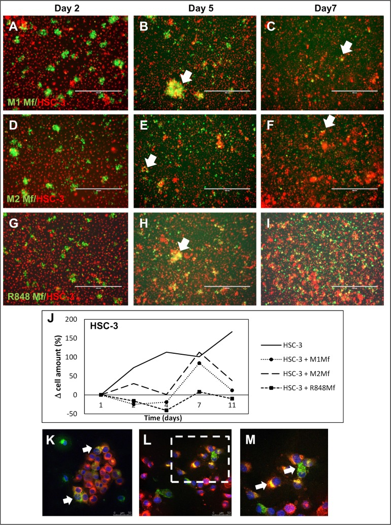
Fig 1. Co-culture of HSC-3 cells and Mfs.
Vybrant CM-Dil labeled HSC-3 (red) and Vybrant DiO labeled Mfs (green) were co-cultured for up to 11 days in normal growth medium and photographed with an Evos FL Cell Imaging System microscope. Cell density was analyzed optically using Leica QWin3 Software. Co-culture of labeled HSC-3 and M1 Mfs (A-C), HSC-3 and M2 Mfs (D-F) and HSC-3 and R848 Mfs (G-I) at days 2, 5 and 7. White arrows shows merged/fused cells. (J) Relative HSC-3 cell density in co-cultures. Scale bars 400 μm and n = 2. Representative confocal microscope pictures of co-cultured HSC-3 and R848 Mfs are shown in K-M. Fusion of membranes was observed between the two cell types (white arrows). An R848 macrophage appears to deliver parts to surrounding HSC-3 cells (L), larger picture in M. Scale bars 50 μm in K,L and 25 μm in M.

