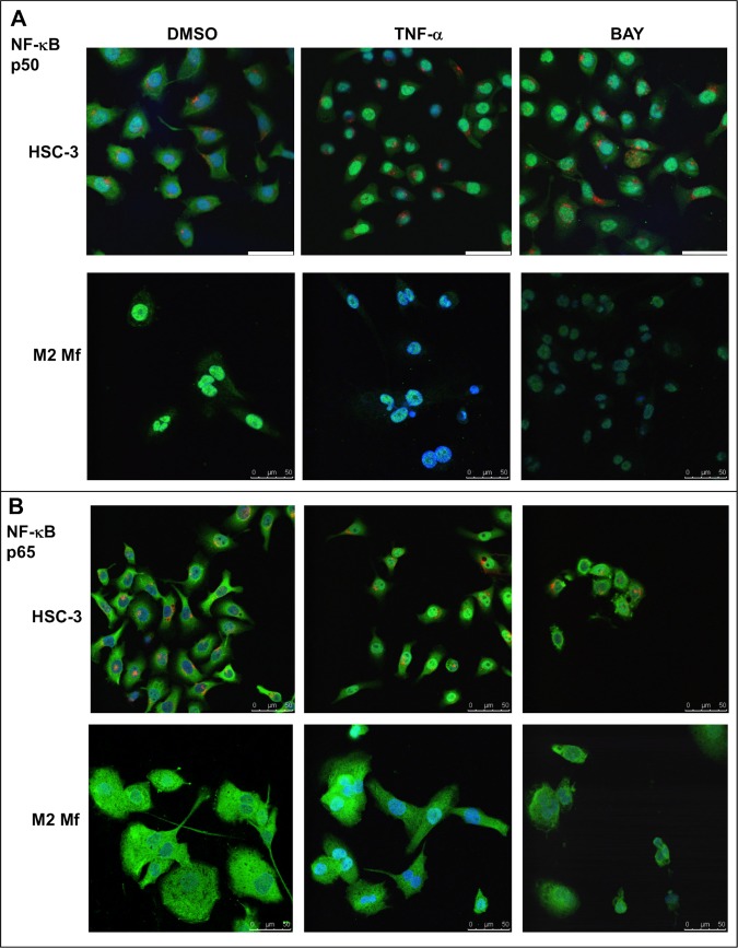
Fig 6. Expression of NF-κB p50 and p65 subunit in HSC-3 and M2 Mfs.
Vybrant CM-Dil labeled HSC-3 cells (red) and unlabeled M2 Mfs were incubated with DMSO or 10 ng/ml TNF-α for 30 min where after cells were fixed for immunofluorescence with polyclonal NF-κB p50 (A) or NF-κB p65 (B) antibodies. Some samples were pre-incubated with 10 μM BAY 11-7082 prior to TNF-α activation. AlexaFluor488-conjugated secondary antibody was used for visualization. Samples were mounted with DAPI- mountain medium to visualize nuclei (blue). Samples were photographed with a Leica Confocal microscope with 63x oil immersion objective. Scale bars 50 μm.

