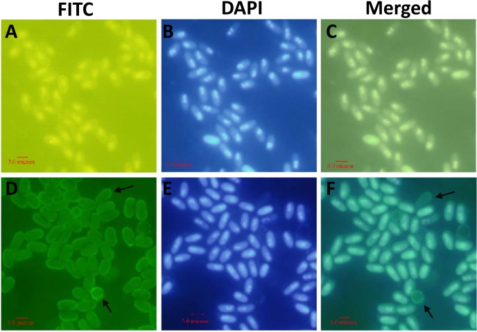
Fig 3. Immunofluorescence labeling of mature spores of N. bombycis.
Spores were permeabilized and detected using mouse IgG (A) and mAb 2B10 (D). The antigen of mAb 2B10, EOB13320, was located on the spore wall. The arrows indicate the empty spore wall. (B) and (E), DAPI staining of the same visual field as in (A) and (D), respectively. (C) and (F), merged images of (A) plus (B) and (D) plus (E), respectively.

