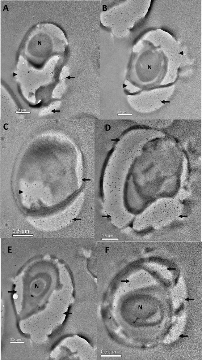
Fig 5. Immunoelectron microscopy of N. bombycis during sporogony.
Samples were prepared and labeled as described in Fig 3. A and B, The developing sporont. The gold labelling is concentrated in the dividing sporoplasm (arrowheads) and the developing endospore (arrows). C-F, Gold labeling was decreased in the sporoplasm and greatly increased in the developing endospore during sporogony (arrows). N, nucleus. Bars = 0.5 μm.

