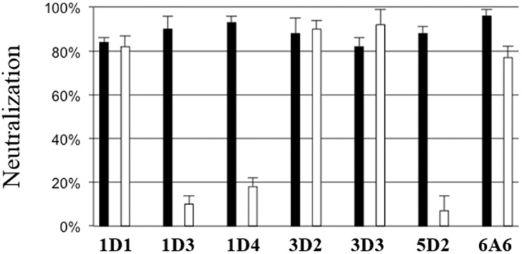
Fig 2. Monoclonal antibodies neutralization mechanism after pre (black) or post (white) MCPyV pseudovirions attachment.

For the detection of neutralizing antibodies, COS-7 cells (104/well) and MCPyV luciferase pseudovirions (0.2 RLU) were used. For the pre attachment determination, pseudovirions were mixed with monoclonal antibodies supernatants diluted 1:3 during 1 h and then added to the cells for 3 h at 37°C. The mixture was removed and 100 μl of DMEM-FCS were added. For investigation of post-attachment neutralization, pseudovirions were bound to cells for 1 h at 4°C. Unbound virions were removed and then antibodies diluted 1:3 were added during 1h. The antibodies were removed and 100 μl of DMEM-FCS were added. After incubation for 48 h at 37°C the luciferase activity was measure. The results were expressed as the percentage of inhibition of luciferase activity. The data presented are the means of three determinations performed in duplicate (+/- SEM).
