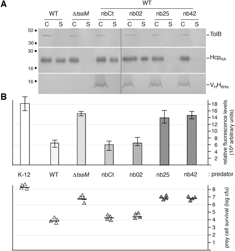
Fig 5. TssM-specific nanobodies nb25 and nb42 specifically affect T6SS function.
(A) Hcp release assay. HA-epitope-tagged Hcp (HcpHA) release was assessed by separating whole cells (C) and supernatant (S) fractions from wild-type (WT), ΔtssM cells or WT cells producing 6His-tagged nanobodies as indicated. 2×10⁸ cells and the TCA-precipitated material of the supernatant from 5×10⁸ cells were loaded on a 12.5%-acrylamide SDS-PAGE and the nanobodies (lower panel), Hcp (middle panel) and periplasmic TolB (cell integrity control, upper panel) proteins were immunodetected using anti-5His, anti-HA and anti-TolB antibodies respectively. (B) Anti-bacterial assay. The Sci-1 T6SS-dependent anti-bacterial activity was assessed by mixing prey cells (W3110 gfp +, kanR) with the indicated attacker cell (K-12, W3110; WT, EAEC 17–2; ΔtssM, 17–2ΔtssM or WT cells producing the indicated nanobody) for 16 hours at 37°C in sci-1-inducing medium (SIM). The recovered fluorescent level (in arbitrary units) is shown in the upper graph (mean of fluorescence levels per OD600nm obtained from four independent experiments). The number of recovered viable prey cells, expressed in colony forming unit (cfu) is shown in the lower graph (the triangles indicate values from four independent assays, and the average is indicated by the bar).

