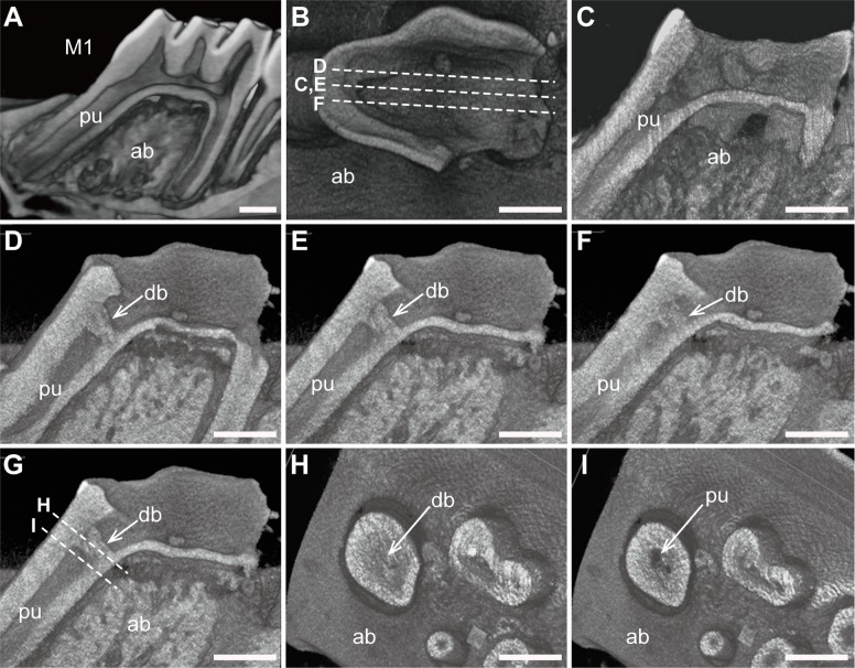
Fig 2. Reconstructed micro-CT images and two-dimensional virtual slices.
(A) Three-dimensional reconstructed image of the intact lower first molars in a 10-week-old rat. (B) Virtual sagittal slices were obtained in the control (C) and experimental groups (D-F). (C) No dentin bridges were observed in any of the control mice. (D-F) Series of reconstructed images showing that LiCl treatment induced dentin bridge formation over the exposed pulp surface. (G) Virtual coronal slices were obtained in the experimental group. (H,I) Reconstructed coronal images also illustrated deposition of reparative tissues (H). Abbreviations: M1, first molars; ab, alveolar bone; pu, pulp; db, dentin bridge. Scale bars, 1 mm.

