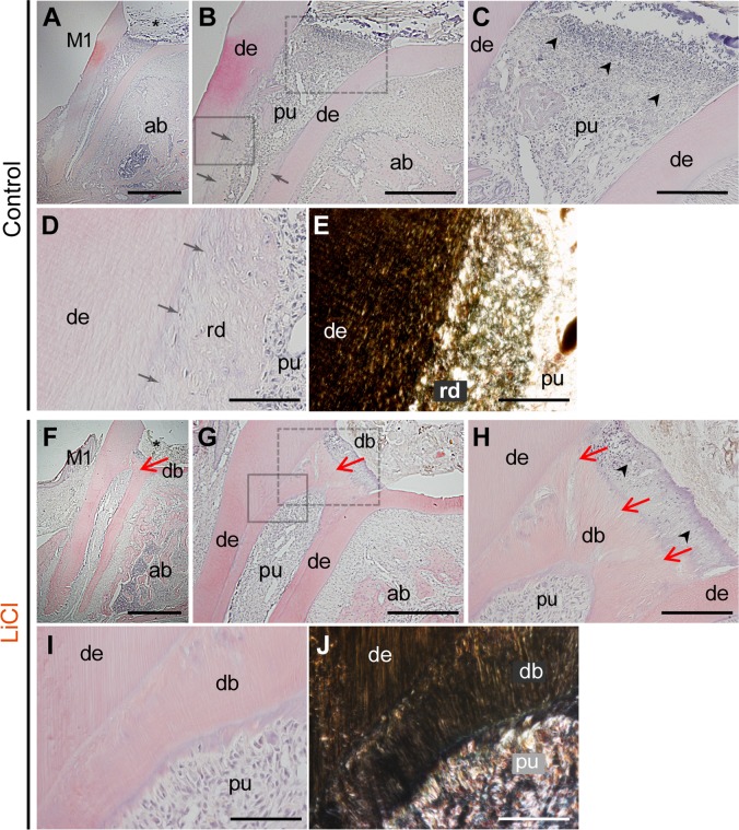
Fig 3. Histological micrographs (H-E) of sagittal sections of the first upper molars.
Reparative dentin formation was evaluated on the serial sections in the control (A-E) and LiCl (F-J) groups at four weeks after pulpotomy. (A,F) Pulpotomy was performed in the first molars (M1), and the coronal part of the pulp was removed (asterisk). (B) In the control molars, no reparative dentin bridges were observed beneath the pulp exposure site. However, hard reparative tissue was detected along the residual root dentin surface (arrows). (C) Higher magnification view of the dot box in panel B. Necrotic and disintegrated tissue was observed just beneath the pulp exposure site (arrow heads). (D,E) Bright (D) and dark (E) field images at higher magnification of the solid box in panel B. Dentin tubules were observed in the residual dentin (de), whereas a tubular structure was hardly detected in the deposited reparative tissue (rd). (F-G) The LiCl group exhibited dentin bridges beneath the exposed surface, and the reparative tissue further expanded in the direction of the apex (red arrows). (H) Higher magnification view of the dot box in panel G. A tubular structure was evident in the dentin bridges (red arrows), with only a few cells entrapped in the matrix. (I,J) Bright (I) and dark (J) field images at higher magnification of the solid box in panel G. The reparative matrix in the LiCl group was more condensed, with a well-developed tubular structure, than that observed in the control group. The figure represented the similar results from independent samples. Abbreviations: M1, the first upper molar; mr, mesial root; ab, alveolar bone; de, dentin; pu, pulp; rd, regenerative dentin, db, dentin bridge. Scale bars, 100 μm in A,F; 50 μm in B,G; 20 μm in C,H; 10 μm in D,E,I,J.

