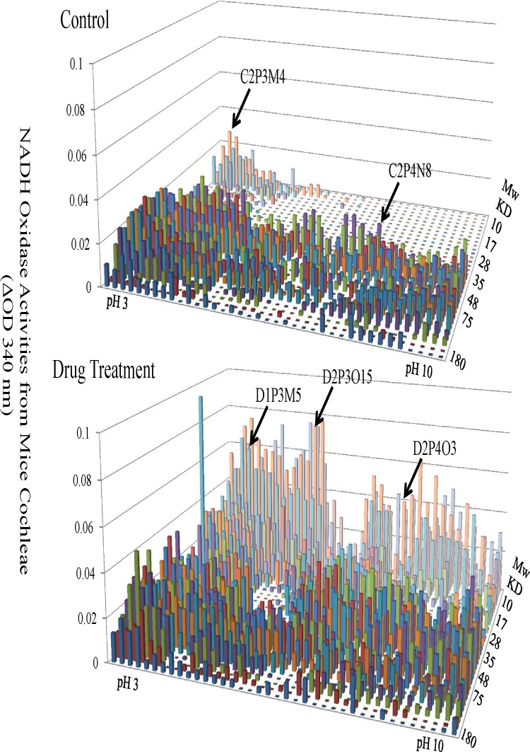
Fig 3. Changes of NADH-dependent Oxidase Activities in Mouse Cochleae from the Control and Drug Treatment Groups.
The functional landscape of NADH-dependent oxidases from mouse cochleae with (bottom panel) or without drug treatment (top panel) were shown. These oxidases were separated by 2-D electrophoresis based on their unique isoelectric points (X axis) and molecular weight (Y axis). Their activities were shown as the ability to oxidize NADH (Z axis).

