
Figure 2. Olfactory systems from 3 phyla.
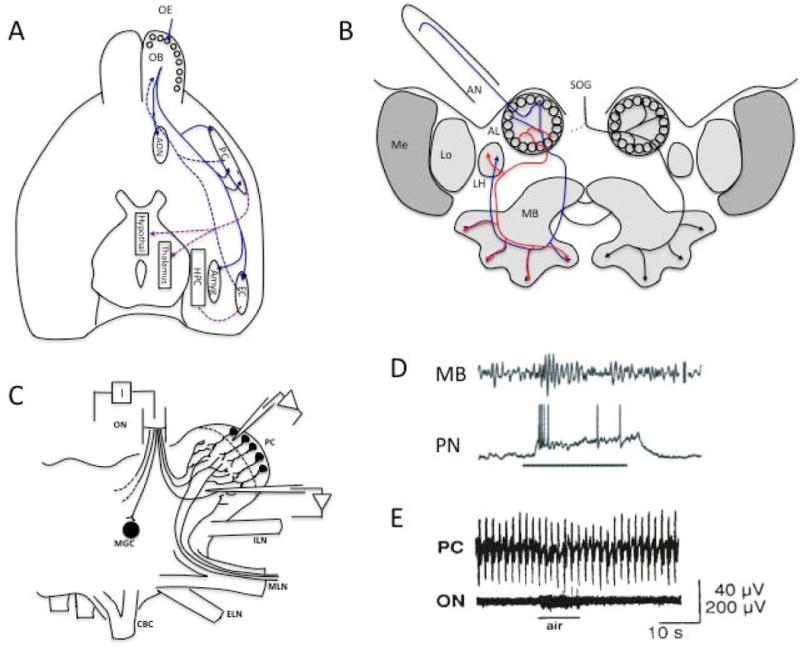
A. The mammalian central olfactory system begins with the OB, which receives olfactory nerve input from the olfactory epithelium (OE) in the glomeruli around the periphery of the bulb. Primary olfactory cortex is primarily represented by the anterior olfactory nucleus (AON) [64]. The pyriform cortex (PC) is a higher order sensory association cortex [65,66], but it is often referred to as primary olfactory cortex. The OB also projects to the multimodal entorhinal cortex (EC), which sends fibers into the hippocampus (HPC), and the amygdala (amyg) among other limbic and subcortical areas. The PC projects to the EC, hypothalamus and thalamus, as well as other higher order areas. Most OB connections to other brain regions are bidirectional. Centrifugal projections to the OB synapse primarily onto GABAergic GCs in the deep layers, except for the AON, which targets superficial juxtaglomerular cells and mitral cells [67]. (Figure adapted from [68].) B. The honeybee central olfactory system begins with antennal nerve (AN) input to the antennal lobe (AL) projection neurons in peripheral glomeruli. AL neurons project to the mushroom body (MB), a higher order multimodal area, and the lateral horn (LH). Descending modulatory input associated with appetitive state comes from the VUM-mx optopaminergic neuron in the subesophogeal ganglion (SOG). (Figure adapted from [69]). C. The limax cerebral ganglion receives olfactory nerve (ON) input to glomeruli in the procerebrum (PC). The PC also receives input from the medial lip nerve (MLN) in the inferior nose. (Figure adapted from [15] with permission.) D. Odor evoked oscillations (~20 Hz) are recorded in the locust mushroom body (MB) but are produced by axon terminals from the antennal lobe (AL) projection neurons (PN). Simultaneously recorded PN shows depolarization, odor evoked spikes and subthreshold oscillations. (Figure adapted from [26], with permission). E. Procerebral lobe oscillations in limax showing the <1Hz oscillation typical of this species. (Figure adapted from [15] with permission.)