
Figure 3. A. Olfactory bulb circuitry associated with oscillations.
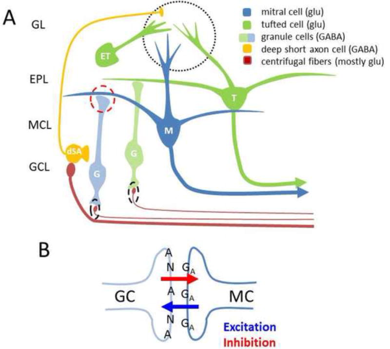
Over the past few years, the canonical picture of OB circuitry has changed and some of the changes may have implications on mechanism and modification of OB oscillations: 1) Olfactory nerve input (not shown) targets external tufted (ET) cells directly (and possibly all tufted cells within glomeruli [31]) and then MT cells via excitatory inputs from ETs and inhibitory relays from ETs to periglomerular (PG) cells (not shown) to MT cells. ETs fire in bursts that can match the respiratory rhythm and may support theta oscillations [70]. One population of GABAergic deep short axon cells (dSAC) target PG cells [36]. Pyriform cortex input targets dSACs [37]. B. Reciprocal synapse as shown in the red dashed circle in A is on the distal dendrites of GCs and may support local graded inhibition through AMPA receptors with ~4.2 ms rise times [17]. This mechanism may support gamma oscillations independent of GC spikes [21]. NMDA and AMPA receptors are present on GCs at most synapses. About 25% are NMDA silent and a very small number are AMPA silent. GABAA receptors mediate inhibition of MTs. Synapses proximal to the GC soma from centrifugal axon fibers also have NMDA and AMPA receptors at most of these synapses (dashed black ovals). Activation of GCs by stimulating these fibers produces GABA release at the reciprocal synapse with faster rise times (1.3 ms). Additional abbreviations: GL- glomerular layer, EPL- external plexiform layer, MCL- mitral cell layer, GCL-granule cell layer.