Abstract
Phrenic long-term facilitation (pLTF) is a form of respiratory plasticity induced by acute intermittent hypoxia (AIH) or episodic carotid chemoafferent neuron activation. Surprisingly, residual pLTF is expressed in carotid denervated rats. However, since carotid denervation eliminates baroreceptor feedback and causes profound hypotension during hypoxia in anesthetized rats, potential contributions of these uncontrolled factors or residual chemoafferent neuron activity to residual pLTF cannot be ruled out. Since ATP is necessary for hypoxic carotid chemotransduction, we tested the hypothesis that functional peripheral chemoreceptor denervation (with intact baroreceptors) via systemic P2X receptor antagonism blocks hypoxic phrenic responses and AIH-induced pLTF in anesthetized rats. Pyridoxal-phosphate-6-azophenyl-2′,4′-disulfonic acid (PPADS; 100 mg/kg i.v.), a non-selective P2X receptor antagonist, was administered to anesthetized, vagotomized, paralyzed and ventilated male Sprague–Dawley rats prior to AIH (3, 5 min episodes of 10% O2; 5 min intervals). Although PPADS strongly attenuated the short-term hypoxic phrenic response (20±4% vs. 113±15% baseline; P < 0.001), pLTF was reduced but not eliminated 60 min post-AIH (25±4% vs. 51±11% baseline; n = 8 and 7, respectively; P < 0.002). Thus, AIH initiates residual pLTF out of proportion to the diminished hypoxic phrenic response and chemoafferent neuron activation. Although the mechanism of residual pLTF following functional chemo-denervation remains unclear, possible mechanisms involving direct effects of hypoxia on the CNS are discussed.
Keywords: Long-term facilitation, Intermittent hypoxia, PPADS, ATP receptor, Carotid chemoreceptor, Plasticity, Motor neuron
1. Introduction
Phrenic long-term facilitation (pLTF) is a widely studied model of respiratory plasticity characterized by a persistent increase in phrenic motor output lasting hours after exposure to acute intermittent hypoxia (AIH) (Mitchell et al., 2001; Mahamed and Mitchell, 2007). Intermittent electrical stimulation of chemoafferent neurons in the carotid sinus nerve is sufficient to elicit pLTF in anesthetized cats (Millhorn et al., 1980a,b) and rats (Hayashi et al., 1993). Since both AIH-induced pLTF and pLTF following carotid chemoafferent activation are serotonin-dependent, they presumably share a common mechanism (Millhorn et al., 1980b; Bach and Mitchell, 1996). On the other hand, carotid denervation reduces the short-term hypoxic phrenic response by ~75% in anesthetized rats, yet robust pLTF is still observed (~50% of normal) (Bavis and Mitchell, 2003). Thus, mechanisms independent from chemoafferent neuron activation may give rise to an attenuated form of pLTF (Bavis and Mitchell, 2003).
Carotid denervation eliminates carotid chemoafferent and carotid baroreceptor input to the central nervous system (CNS). Thus, it is difficult to conclude if responses post-carotid denervation involve loss of chemoreceptor versus baroreceptor feedback. Carotid denervation exacerbates hypoxia-induced hypotension in anesthetized rats (Bavis and Mitchell, 2003), largely due to the loss of baroreflexes and carotid chemoafferent inputs to C1 cardioregulatory neurons of the ventrolateral medulla (Aicher et al., 1996; Koshiya and Guyenet, 1996; Paton et al., 2001). Greater hypotension during hypoxic episodes may lead to greater CNS hypoxia, potentially inducing alternate mechanisms of AIH-induced plasticity. Thus, it is difficult to interpret residual pLTF in anesthetized rats following surgical carotid denervation. Here we utilize a novel, functional (chemical) denervation of carotid chemoafferent neurons to re-investigate the need for chemo-afferent neurons in pLTF.
ATP, acting via ionotropic P2X2 receptors, is a critical neurotransmitter in peripheral chemoreceptor function (Prasad et al., 2001; Rong et al., 2001; Zhang et al., 2000). As such, ATP receptor antagonists that do not cross the blood–brain barrier inhibit carotid chemoafferent activity during hypoxia, while maintaining chemoafferent synaptic connections in the CNS. Thus, ATP receptor antagonists elicit temporary and reversible attenuation of chemoafferent activity. We utilized systemic application of a non-selective P2X receptor antagonist (PPADS) to block peripheral chemosensory transduction while leaving the carotid sinus nerve (and baroreceptor function) intact. Our original hypothesis was that systemic PPADS administration would block the short-term hypoxic phrenic response and AIH-induced pLTF. In contrast, although the short-term hypoxic response was nearly abolished (<20% of normal), residual AIH-induced pLTF was still observed (>50% of normal). Thus, hypoxia may contribute to AIH-induced pLTF independently from peripheral chemoreceptor activation, possibly via direct effects on the CNS.
2. Methods
Experiments were conducted on 3–5-month-old male Sprague–Dawley rats (300–350 g; Harlan 218a; Harlan, Indianapolis, IN). Animals were doubly housed in a controlled environment (12 h light/dark cycle) with food and water available ad libitum. The Animal Care and Use Committee at the University of Wisconsin approved all protocols.
2.1. Experimental preparation
Anesthesia was induced with isoflurane and maintained (3.0–3.5% isoflurane, FIO2 = 0.5, balance N2) through a nose cone. After tracheal cannulation to allow pump ventilation (Tidal volume 2.5 ml; Rodent Respirator model 682; Harvard Apparatus, South Natick, MA), isoflurane was continued via the ventilator. End-expiratory carbon dioxide partial pressures (PETCO2) were continuously monitored with a CO2 analyzer in a flow-through configuration (Capnogard; Novametrix Medical Systems, Wallingford, CT). Animals were bilaterally vagotomized in the mid-cervical region to prevent phrenic nerve entrainment with the ventilator. The right femoral artery was cannulated to monitor blood pressure (P23ID pressure transducer and P122 amplifier, Gould) and to withdraw 0.2 ml blood samples for blood–gas analysis (ABL-500, Radiometer, Copenhagen, Denmark). A 24-gauge catheter was placed in a tail vein for administration of pharmacologic agents and continuous intravenous fluids (5 ml/kg/h; 50:50 mixture of lactated Ringer’s and 6% Hetastarch). Body temperature was monitored and maintained at 37.0±1 °C via placement of a rectal probe and a heated surgical table. The left phrenic nerve was isolated via a dorsal approach, cut distally, de-sheathed, submerged in mineral oil and placed on bipolar silver wire electrodes. Upon completion of surgical preparation, animals were converted to urethane anesthesia (1.8 g/kg, i.v.) and paralyzed with pancuronium bromide (2.5 mg/kg, i.v.) to prevent spontaneous breathing movements.
2.2. Experimental protocol
The CO2 apneic threshold was determined by decreasing inspired CO2 and/or increasing ventilator frequency until phrenic minute activity ceased for >60 s. Subsequently, the CO2 recruitment threshold was determined by increasing inspired CO2 and/or decreasing ventilator frequency until phrenic activity resumed. Baseline phrenic activity was standardized between animals by maintaining PETCO2 2–3mmHg above the recruitment threshold. Rats were then administered either pyridoxal-phosphate-6-azophenyl-2′,4′-disulfonic acid (PPADS), a non-selective P2X receptor antagonist (100 mg/kg, i.v.; 10 mg/ml in 0.9% NaCl; n = 14), or vehicle (0.9% NaCl; 10 ml/kg, i.v.; n = 13). PPADS elicited an increase in integrated phrenic burst amplitude associated with an increase in PaCO2, despite constant PETCO2 . Because of the increased PETCO2 to PaCO2 difference, we lowered PETCO2 in some experiments until phrenic activity matched pre-drug levels; at this point, PaCO2 matched the original baseline value. Since this adjustment was performed only in half of the PPADS-treated rats (each group n = 4), they were divided into ventilation-adjusted and non-adjusted groups. However, in subsequent statistical comparisons between these groups, no differences were detected; thus, in all data presentations, these groups were combined into a single control group.
Phrenic neural output was allowed to stabilize over 15 min following drug or vehicle administration. Subsequent analysis of arterial blood (0.2–0.3 ml in heparinized glass tuberculin syringe) characterized baseline PaCO2, PaO2, pH and base excess values. Rats were then exposed to either acute intermittent hypoxia (3, 5min bouts of hypoxia; PaO2 =40±5mmHg; n = 15) or maintained under baseline oxygen conditions (time controls; n = 12). Arterial blood gases were analyzed at 15, 30 and 60 min following AIH (or at equivalent times in control experiments) to confirm isocapnia with respect to baseline values (±1.5mmHg baseline PaCO2). Upon completion of this protocol, rats were euthanized via an overdose of urethane.
A total of 27 animals were used in this study, with experimental groups were divided as follows: 14 animals were administered PPADS, 8/14 were exposed to AIH and 6/14 were PPADS time controls. Thirteen animals were administered vehicle (saline); 7/13 were exposed to AIH and 6/13 were vehicle time controls.
2.3. Electrophysiological data analysis
Nerve activity was amplified (10,000×), band-pass filtered (100 Hz to 10 kHz; Model 1700 or 1800, A-M Systems, Carlsborg, WA), and integrated (time constant = 50ms; Moving Averager, model MA-821RSP, CWE, Ardmore, PA). The integrated signal was digitized and processed with commercially available computer software (WinDaq 2.18, DATAQ Instruments, Akron, OH).
Peak integrated phrenic amplitude, phrenic burst frequency and mean arterial blood pressure were averaged in 240 s bins (immediately preceding blood sampling) under baseline conditions, 15, 30 and 60 min following the final hypoxic episode. A 60-s bin during the fifth minute of hypoxia, or equivalent time point during baseline conditions, was also averaged. Burst amplitude and phrenic minute activity were normalized as a percentage change from baseline activity; frequency was expressed as a change from baseline in bursts/min. Data were compared using a two-way repeated measures ANOVA followed by a Student–Newman–Keuls post hoc test. Short-term hypoxic responses, PaCO2 vs. PETCO2 differences and mean arterial pressure differences were analyzed using unpaired t-tests. Linear regression analysis was performed to assess correlations between changes in mean arterial pressure and pLTF. Effects were considered statistically significant at P≤0.05. Statistical tests were conducted via SigmaStat 2.03, SPSS, Chicago, IL. All data are represented as means±SEM.
3. Results
3.1. Baseline physiologic effects of PPADS
Upon systemic PPADS administration, multiple physiological changes were observed. Mean arterial pressure (MAP) during baseline conditions was significantly increased from vehicle-treated rats (130±20 vs. 110±6, n = 14 and 13, respectively; P = 0.02). Following drug administration, a slight apparent decrease in PaO2 was observed (261±13mmHg vs. 202±24mmHg, pre- and post-drug, respectively; P = 0.08), accompanied by a significant elevation in PaCO2 (10±2mmHg; P < 0.001). Thus, it was necessary to adjust PETCO2 at a lower level post-drug administration (45±1mmHg vs. 39±3mmHg) to attain a constant level of PaCO2 (47±1mmHg vs. 47±2mmHg, each n=4; P = 0.9). PPADS initially increased phrenic motor output via increases in PaCO2 since phrenic motor output returned completely to initial baseline values once baseline PaCO2 values had been restored.
3.2. Time control experiments
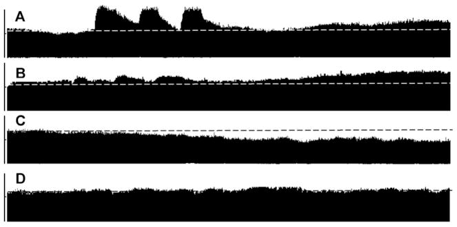
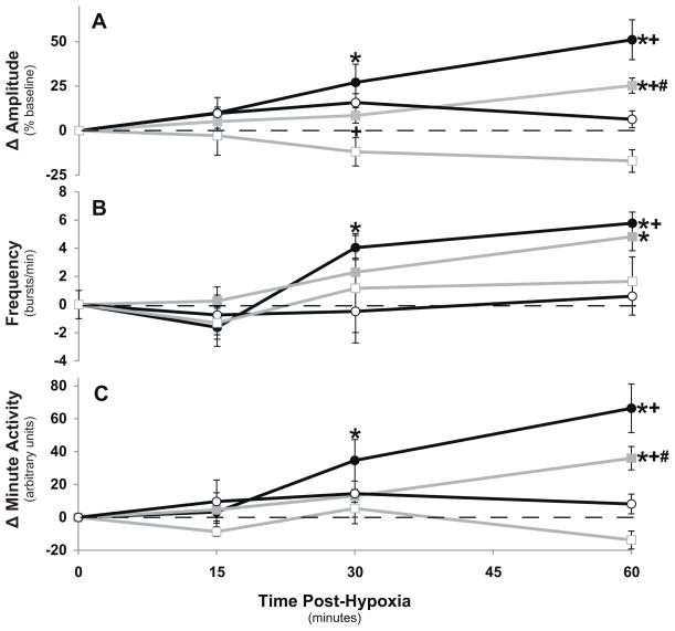
Vehicle and PPADS time control experiments exhibited no statistically significant differences in phrenic activity from baseline at any time during the protocol (Figs. 1 and 3). There was a trend for decreasing peak integrated phrenic burst amplitude in PPADS-treated rats, but this trend was not significant (60 min: −17±6% baseline; P = 0.1; n = 6).
Fig. 1.

Representative tracings of compressed phrenic nerve recordings during experimental protocols. (A) AIH following vehicle injections, demonstrating normal pLTF. (B) AIH with PPADS, demonstrating residual pLTF. (C) Time control experiment following PPADS administration without AIH. (D) Time control experiment following vehicle injection without AIH. Vertical bars to the left of each experimental trace represent 1V. Dotted horizontal lines represent the initial, baseline value of phrenic burst amplitude. The time represented after the final hypoxic episode is 1 h.
Fig. 3.
Changes in phrenic activity from baseline after episodic hypoxia in vehicle-treated (●, n = 7) and PPADS-treated (■, n = 8) rats. Time control rats without AIH are also shown (vehicle-treated, ○, n = 6; PPADS-treated, □, n = 6): (A) phrenic burst amplitude; (B) phrenic burst frequency; (C) minute phrenic activity. Values are means±1 SEM. *Different from baseline, P < 0.005; +different from corresponding time control group (i.e. same drug), P < 0.005; #different from corresponding vehicle control group (i.e. same protocol, AIH or hyperoxia) at same time point, P < 0.005.
3.3. Effects of PPADS on short-term hypoxic phrenic responses (STHPR)
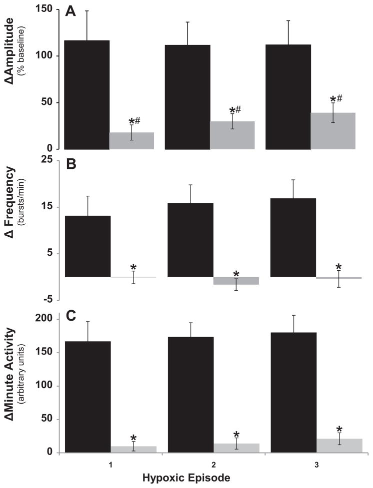
Each of the 3 hypoxic episodes increased phrenic burst amplitude in vehicle-treated rats, and there were no significant differences in response among episodes (116±32, 112±25, and 112±26% baseline, n=7; P = 0.99). PPADS significantly attenuated the STHPR (Fig. 2A, n=8; P < 0.001). Despite an apparent trend, there was no significant difference among the 3 AIH-induced STHPRs in PPADS-treated rats (13±5, 21±7, and 26±7% baseline; P = 0.35) indicating that drug effects persisted throughout the AIH exposure. On average, integrated phrenic responses following PPADS were less than 18% of control rats (113±15% vs. 20±4% baseline for control and PPADS treated rats, respectively; P < 0.001).
Fig. 2.
Changes in phrenic burst amplutude (A), burst frequency (B) and minute phrenic activity (C) during each of three hypoxic episodes (i.e. AIH) in vehicle-treated (filled bars, n = 7) and PPADS-treated (open bars, n = 8) rats. Values are means±1 SEM. *Different from baseline, P < 0.005; #different from corresponding vehicle control (i.e. same protocol, AIH or hyperoxia), P < 0.05.
Burst frequency increased during hypoxic episodes in vehicle-treated rats (Fig. 2B), and there was no difference in response among episodes (13±4, 16±4, and 17±4 bursts/min; P = 0.8). However, there was no change in burst frequency during hypoxic episodes following PPADS administration (0±1, −2±1, and 0±2 bursts/min; Fig. 2B; P = 0.21). Thus, PPADS abolished increases in burst frequency during hypoxia (−0.6±1 vs. 15±9 for PPADS and control, respectively; P < 0.001). Accordingly, phrenic minute activity during hypoxic episodes was significantly attenuated relative to control rats (Fig. 2C); the phrenic minute activity response during hypoxia was ~15% that of control rats following PPADS administration (144±15% vs. 21±4% baseline; P < 0.001).
3.4. Phrenic long-term facilitation
AIH exposed vehicle control rats exhibited significant increases in phrenic burst amplitude at 30 and 60 min post-AIH versus comparable times in control experiments without AIH, thus demonstrating pLTF (30 min: 27±10%; 60 min: 51±11% baseline; P < 0.001). Phrenic burst frequency also increased from baseline at 30 and 60 min post-AIH (4.0±1 and 5.8±0.8 bursts/min, respectively; P < 0.05; Fig. 3B), although this effect is much smaller on a percentage basis, consistent with other reports using this experimental preparation (Baker-Herman and Mitchell, 2008). Accordingly, phrenic minute activity in vehicle-treated rats was increased from baseline at 30 and 60 min post-AIH (35±3 and 66±5% baseline, respectively; P < 0.001).
Despite substantially attenuated STHPRs, AIH elicited significant pLTF in PPADS-treated rats (P < 0.001). Following AIH, phrenic amplitude progressively increased, resulting in a significant increase from baseline by 60 min post-AIH in PPADS treated rats (25±4%; P = 0.002; Fig. 3A). This response was significantly attenuated compared to vehicle control rats at 60 min post-AIH (25% vs. 51% baseline, P < 0.001). Phrenic burst frequency also increased from baseline at 60 min post-AIH in PPADS-treated rats (4.8±1.2 bursts/min; P = 0.002; Fig. 3B), but this response was not significantly different from vehicle-control rats. Phrenic minute activity was significantly increased from baseline 60 min post-AIH in PPADS-treated rats (36.0±7.1%; P < 0.001; Fig. 3C), although this response was significantly attenuated versus vehicle control rats.
3.5. Blood pressure regulation
Hypoxia elicited hypotension in both vehicle and PPADS-treated rats (P < 0.05). However, the drop in mean arterial pressure (MAP) from baseline during hypoxia was greater in PPADS-treated rats (−64±6mmHg vs. −19±4mmHg; P < 0.001). MAP also decreased significantly more from baseline at 60 min post-AIH in PPADS-treated versus vehicle control rats (−33±5 vs. −16±4; P = 0.02). Despite a greater change from baseline values, absolute MAP values were not different between PPADS and vehicle-treated rats 60 min post-AIH (97±7mmHgvs. 97±7mmHg; P = 0.9) due to the greater initial value of PPADS treated rats.
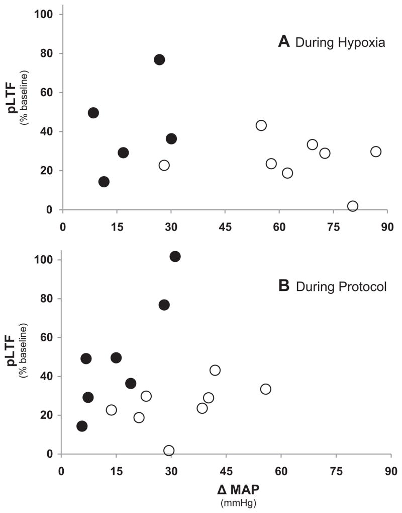
Regression analyses were performed to determine if individual pLTF values correlated with the decrease in MAP during hypoxia (Fig. 4A) or with the decrease in MAP throughout an experimental protocol (Fig. 4B). There was no significant correlation between the change in MAP during hypoxia and pLTF (P = 0.24, r2 = 0.124) in vehicle-treated (n = 7) or PPADS-treated (n = 8) rats. Similarly MAP at 60min post-AIH and pLTF are not correlated (P = 0.72, r2 = 0.01) in PPADS-treated rats. However, a significant correlation was observed between pLTF and MAP at 60min post-AIH in vehicle-treated rats (P = 0.01, r2 = 0.72). This observation is not consistent with numerous previous studies from our laboratory (Fuller et al., 2000; Baker-Herman and Mitchell, 2008), and we regard it as a spurious result here.
Fig. 4.
Regression analysis of pLTF magnitude vs. the change in mean arterial pressure during hypoxia (A), or the change in mean arterial pressure during an experimental protocol (B; 60 min post-AIH vs. baseline). In (A), there was no significant correlation between the change in MAP during hypoxia and pLTF (P = 0.24, r2 = 0.124) in vehicle-treated (●, n = 7) and/or PPADS-treated (○, n = 8) rats. Similarly (B) MAP at 60 min post-AIH and pLTF are not significantly correlated (P = 0.72, r2 = 0.01) in PPADS-treated rats. However, a significant correlation was apparent between pLTF and MAP at 60min post-AIH in vehicle-treated rats (P = 0.01, r2 = 0.717). This latter observation is not consistent with numerous previous observations from our laboratory and is regarded as a spurious result here (see discussion; Baker-Herman and Mitchell, 2008).
Similar regression analyses between the magnitude of the STHPR and pLTF revealed no significant correlation (P = 0.79; data not shown). However, a significant correlation was observed between the drop in MAP during hypoxia and the STHPR (P = 0.04; r = 0.57; data not shown). This finding is consistent with predictions that pLTF is independent of the STHPR in PPADS treated rats. On the other hand, it suggests a relationship between the STHPR and the drop in MAP, consistent with the idea that loss of a common factor (e.g. chemoafferent neuron activity) at least partially underlies both responses.
4. Discussion
The relative contributions of peripheral chemoafferent neurons versus baroreceptors and/or (brain)tissue hypoxia to AIH-induced pLTF were investigated by functional chemical denervation of peripheral chemoreceptors via systemic P2X ATP receptor antagonism. Systemic application of the P2X receptor antagonist PPADS greatly attenuates the short-term hypoxic phrenic response (<20% of normal), but diminishes pLTF to a lesser extent, suggesting that mechanisms capable of eliciting pLTF persist without (or with minimal) chemoafferent neuron activation. In the absence of functional chemoafferent neurons, other mechanisms may substitute for the normal, serotonin-dependent mechanism of pLTF. We speculate that severe CNS hypoxia caused by combined hypoxemia and hypotension during AIH in PPADS treated rats elicits a novel adenosine-dependent mechanism of phrenic motor facilitation.
4.1. PPADS effects on physiological variables
4.1.1. Blood gas regulation
Systemic PPADS elicited a brisk increase in phrenic activity immediately following drug administration, an effect that persisted throughout the baseline period. This effect is consistent with the observed PPADS-induced hypercapnia, which can be explained by known effects of ATP receptors on pulmonary circulation (Burnstock, 2007) and subsequent effects on ventilation-perfusion relationships in the lung. At a given level of (pump-controlled) ventilation, disruptions in ventilation–perfusion relationships are expected to reduce the efficiency of gas exchange, causing hypoxemia and hypercapnia. However, the hypoxemia was offset by elevated inspired oxygen levels used throughout our experiments. In some rats, adjustments were made, reversing the hypercapnia and completely restoring baseline phrenic nerve activity (prevs. post-PPADS). This adjustment had no consistent effect on the fundamental conclusions of this study concerning either the short-term hypoxic response or pLTF.
4.1.2. Mean arterial pressure (MAP)
PPADS significantly increased MAP (20mmHg; P = 0.017), contrary to reports that ATP receptor activation increases cardiac contractility and systemic vascular resistance (Burnstock, 2007). PPADS-induced hypertension persisted throughout baseline measurements. ATP also induces bradycardia and hypotension via P2X receptor activation in the ventro-lateral medulla (Thomas et al., 2001). Since there is no available evidence to suggest that PPADS crosses the blood–brain barrier, PPADS may exert previously undescribed vasoactive effects in rats.
The 3-fold greater decrease in MAP during hypoxia in PPADS-treated rats may reflect inhibition of chemoreflex pressor responses (i.e. chemoafferent neurons no longer mitigate the effects of hypoxic vasodilation) (Aicher et al., 1996; Kara et al., 2003; Olson et al., 2001). The hypotension may be exacerbated due to more severe tissue hypoxia, greater ATP release and greater adenosine accumulation, thereby inducing greater vasodilation. Greater CNS tissue hypoxia and adenosine accumulation may elicit pLTF by a novel adenosine-dependent mechanism associated with TrkB trans-activation versus new BDNF synthesis (Golder et al., 2008). However, the lack of correlation between decreased MAP during hypoxia and pLTF (Fig. 4) may argue against this hypothesis.
The greater decrease in MAP from baseline to the 60 min post-AIH time point in PPADS treated rats may have arisen from progressively diminishing PPADS levels and its hypertensive effects. However, the lack of correlation between the drop in blood pressure and pLTF at 60 min post-AIH suggest that MAP changes throughout the protocol did not cause residual pLTF, at least not in any simple way.
4.2. Phrenic responses
4.2.1. Short-term hypoxic phrenic response
Carotid chemotransduction is critically dependent upon ATP and (specifically) P2X2 receptor activation (Gourine et al., 2005; Prasad et al., 2001; Rong et al., 2001; Zhang et al., 2000). PPADS attenuated the short-term hypoxic phrenic response by >80%, confirming ATP’s critical role in peripheral chemoreflexes. The residual hypoxic response consisted of changes in peak integrated phrenic amplitude, without changes in phrenic burst frequency, an effect consistent with (surgically) carotid-denervated rats (Bavis and Mitchell, 2003). However, the short-term hypoxic response was not completely abolished by PPADS, suggesting the drug may not have been completely effective, or that there is extra-carotid hypoxic chemosensation. In two preliminary studies using lower PPADS doses, identical suppression of the short-term hypoxic phrenic response was observed (data not shown). Thus PPADS doses were most likely sufficient to elicit maximal effects, suggesting that residual hypoxic phrenic responses arise from other (non-PPADS sensitive) mechanisms. The possibility of non-carotid peripheral chemoreceptors, such as aortic and abdominal receptors (Brophy et al., 1999; Martin-Body et al., 1985), is unlikely since the bilateral vagotomy performed in this preparation eliminates the afferent pathways of these alternative peripheral chemoreceptors. Since acetylcholine is a necessary co-transmitter in carotid chemotransduction (Zhang et al., 2000), ACh may mediate residual effects in this study since Ach receptors were not targeted by PPADS. On the other hand, a third, more likely possibility is that residual STHPR results from hypoxic effects in the CNS. Residual hypoxic responses are observed following carotid-denervation in awake and anesthetized rats (Bavis and Mitchell, 2003; Martin-Body et al., 1985). Similar respiratory patterns during the residual hypoxic response in carotid-denervated and PPADS-treated rats are consistent with the conclusion that systemic PPADS effectively induced functional (chemical) carotid denervation. We suggest that the small, residual STHPR most likely arises from hypoxic effects on the CNS.
4.2.2. Phrenic long-term facilitation
Acute intermittent hypoxia induced robust pLTF despite substantial attenuation of the hypoxic phrenic response (~18%), similar to carotid chemodenervated rats (Bavis and Mitchell, 2003). Although increased phrenic motor output following AIH in vehicle and PPADS-treated rats is predominantly due to increased amplitude, a modest frequency facilitation was also observed in both groups.
The significant correlation between phrenic burst amplitude and changes in MAP in vehicle-treated rats at 60 min post-AIH is not consistent with many previous studies from our laboratory using this same experimental preparation; extensive meta-analyses indicate that there is no correlation between post-AIH changes in MAP and pLTF magnitude (Fuller et al., 2000; Baker-Herman and Mitchell, 2008). Further, experimental decreases of mean arterial pressure via hemorrhage have no significant effect on LTF magnitude (Neverova et al., 2007). Thus, we conclude that pLTF in vehicle-treated rats in this experiment is not dependent on changes in mean arterial pressure and that the observed correlation was a spurious result.
Phrenic burst frequency facilitation is variable, but has been demonstrated without carotid chemoafferent activity during hypoxia (Bavis and Mitchell, 2003; Tadjalli et al., 2007; this study; see Baker-Herman and Mitchell, 2008). ATP release during hypoxia may increase rhythmogenic activity in ATP-sensitive preBotzinger complex neurons (Lorier et al., 2008), potentially contributing to both residual hypoxic phrenic frequency responses and frequency LTF.
Although the mechanism of residual pLTF cannot be established from this study, it may result from continued expression of normal LTF mechanisms or a unique pathway operative when carotid chemoreceptors are absent. Electrical stimulation of carotid chemoafferent axons or arterial hypoxemia both induce pLTF by a mechanism that requires activation of raphe serotonergic neurons (Bach and Mitchell, 1996; Millhorn et al., 1980b). AIH in carotid body intact rats elicits pLTF via spinal 5-HT2 receptor activation (Baker-Herman and Mitchell, 2002; Fuller et al., 2001), new BDNF synthesis (Baker-Herman et al., 2004) and ERK MAP kinase activation (Hoffman and Mitchell, unpublished observations). At present, there is no evidence that pLTF elicited by CNS hypoxia in carotid denervated rats results from similar cellular mechanisms.
Hypoxia increases extracellular levels of ATP and adenosine as ATP is degraded by ecto-nucleotidases (Martín et al., 2007; Parkinson et al., 2005; Phillis et al., 1993; Wallman-Johansson and Fredholm, 1994). Adenosine receptor activation via spinal application of the adenosine 2A receptor agonist, CGS 216880, elicits long-lasting phrenic motor facilitation via a unique mechanism: transactivation of an immature isoform of the high affinity BDNF receptor, TrkB (Golder et al., 2008). This form of phrenic motor facilitation does not require BDNF synthesis, suggesting a distinct mechanism from serotonin-dependent, AIH-induced pLTF in intact rats. Indeed, our data are consistent with the hypothesis that residual pLTF in PPADS treated rats results from AIH-induced adenosine receptor activation. In contrast with this hypothesis, when an adenosine 2A receptor antagonist is applied to normal rats, AIH-induced pLTF is amplified versus diminished (Hoffman et al., 2010). Regardless, AIH contributes to pLTF independently from carotid chemoafferent activity, presumably through direct effects of hypoxia in the CNS.
In summary, we confirm that ATP (via ionotropic P2X receptors) is a critical neurotransmitter in peripheral chemoreceptor function. Although systemic ATP receptor antagonism nearly abolishes the short-term hypoxic phrenic response, robust residual pLTF is observed. Thus, AIH elicits a form of pLTF independently from carotid chemoafferent neuron activity, most likely through direct effects of hypoxia in the CNS.
Acknowledgments
Supported by the National Institutes of Health (HL80209 and T32RR17503). We thank B. Wathen for assistance in preparing the figures and S. Mahamed for assistance with data management.
References
- Aicher SA, Saravay RH, Cravo S, Jeske I, Morrison SF, Reis DJ, Milner TA. Monosynaptic projections from the nucleus tractus solitarii to C1 adrenergic neurons in the rostral ventrolateral medulla: comparison with input from the caudal ventrolateral medulla. J Comp Neurol. 1996;373 (1):62–75. doi: 10.1002/(SICI)1096-9861(19960909)373:1<62::AID-CNE6>3.0.CO;2-B. [DOI] [PubMed] [Google Scholar]
- Bach KB, Mitchell GS. Hypoxia-induced long-term facilitation of respiratory activity is serotonin dependent. Respir Physiol. 1996;104:251–260. doi: 10.1016/0034-5687(96)00017-5. [DOI] [PubMed] [Google Scholar]
- Baker-Herman TL, Mitchell GS. Phrenic long-term facilitation requires spinal serotonin receptor activation and protein synthesis. J Neurosci. 2002;22:6229–6246. doi: 10.1523/JNEUROSCI.22-14-06239.2002. [DOI] [PMC free article] [PubMed] [Google Scholar]
- Baker-Herman TL, Fuller DD, Bavis RW, Zabka AG, Golder FJ, Doperalski NJ, Johnson RA, Watters JJ, Mitchell GS. BDNF is necessary and sufficient for spinal respiratory plasticity following intermittent hypoxia. Nature Neuroscience. 2004;7:48–55. doi: 10.1038/nn1166. (Featured in News and Views, Nature Medicine 10:25–26) [DOI] [PubMed] [Google Scholar]
- Baker-Herman TL, Mitchell GS. Determinants of frequency long-term facilitation following acute intermittent hypoxia in vagotomized rats. Respir Physiol Neurobiol. 2008;162:8–17. doi: 10.1016/j.resp.2008.03.005. [DOI] [PMC free article] [PubMed] [Google Scholar]
- Bavis RW, Mitchell GS. Intermittent hypoxia induces phrenic long-term facilitation in carotid denervated rats. J Appl Physiol. 2003;94:399–409. doi: 10.1152/japplphysiol.00374.2002. [DOI] [PubMed] [Google Scholar]
- Brophy S, Ford TW, Carey M, Jones JFX. Activity of aortic chemoreceptors in the aneasthetized rat. J Appl Physiol. 1999;514:821–828. doi: 10.1111/j.1469-7793.1999.821ad.x. [DOI] [PMC free article] [PubMed] [Google Scholar]
- Burnstock G. Physiology and pathophysiology of purinergic neurotransmission. Physiol Rev. 2007;87:659–797. doi: 10.1152/physrev.00043.2006. [DOI] [PubMed] [Google Scholar]
- Fuller DD, Bach KB, Baker TL, Kinkead R, Mitchell GS. Long term facilitation of phrenic motor output. Respir Physiol. 2000;121:135–146. doi: 10.1016/s0034-5687(00)00124-9. [DOI] [PubMed] [Google Scholar]
- Fuller DD, Zabka AG, Baker TL, Mitchell GS. Physiological and genomic consequences of intermittent hypoxia. Selected contribution Phrenic long-term facilitation requires 5-HT receptor activation during but not following episodic hypoxia. J Appl Physiol. 2001;90:2001–2006. doi: 10.1152/jappl.2001.90.5.2001. [DOI] [PubMed] [Google Scholar]
- Golder FJ, Ranganathan L, Satriotomo I, Hoffman M, Lovett-Barr MR, Watters JJ, Baker-Herman TL, Mitchell GS. Spinal adenosine A2a receptor activation elicits long-lasting phrenic motor facilitation. J Neurosci. 2008;28:2033–2042. doi: 10.1523/JNEUROSCI.3570-07.2008. [DOI] [PMC free article] [PubMed] [Google Scholar]
- Gourine AV, Llaudet E, Dale N, Spyer KM. Release of ATP in the ventral medulla during hypoxia in rats: role in hypoxic ventilatory response. J Neurosci. 2005;25 (5):1211–1218. doi: 10.1523/JNEUROSCI.3763-04.2005. [DOI] [PMC free article] [PubMed] [Google Scholar]
- Hayashi F, Coles SK, Bach KB, Mitchell GS, McCrimmon DR. Time-dependent phrenic nerve responses to carotid afferent activation: intact vs. decerebellate rats. Am J Physiol Regul Integr Comp Physiol. 1993;265:R811–R819. doi: 10.1152/ajpregu.1993.265.4.R811. [DOI] [PubMed] [Google Scholar]
- Hoffman MS, Golder FJ, Mahamed S, Mitchell GS. Spinal adenosine 2A receptor inhibition enhances phrenic long term facilitation following acute intermittent hypoxia. J Physiol (London) 2010;588:255–266. doi: 10.1113/jphysiol.2009.180075. [DOI] [PMC free article] [PubMed] [Google Scholar]
- Kara T, Narkiewicz K, Somers VK. Chemoreflexes—physiology and clinical implications. Acta Physiol Scand. 2003;177 (3):37–384. doi: 10.1046/j.1365-201X.2003.01083.x. [DOI] [PubMed] [Google Scholar]
- Koshiya N, Guyenet PG. NTS neurons with carotid chemoreceptor inputs arborize in the rostral ventrolateral medulla. Am J Physiol. 1996;270 (6 Pt 2):R1273–R1278. doi: 10.1152/ajpregu.1996.270.6.R1273. [DOI] [PubMed] [Google Scholar]
- Lorier AR, Lipski J, Housley GD, Greer JJ, Funk GD. ATP sensitivity of preBötzinger complex neurons in neonatal rat in vitro: mechanism underlying a P2 receptor-mediated increase in inspiratory frequency. J Physiol. 2008;586 (5):1429–1446. doi: 10.1113/jphysiol.2007.143024. [DOI] [PMC free article] [PubMed] [Google Scholar]
- Mahamed S, Mitchell GS. Is there a link between intermittent hypoxia-induced plasticity and obstructive sleep apnoea? Exp Physiol. 2007;92 (1):27–37. doi: 10.1113/expphysiol.2006.033720. [DOI] [PubMed] [Google Scholar]
- Martín ED, Fernández M, Perea G, Pascual O, Haydon PG, Araque A, Ceña V. Adenosine released by astrocytes contributes to hypoxia-induced modulation of synaptic transmission. Glia. 2007;55:36–45. doi: 10.1002/glia.20431. [DOI] [PubMed] [Google Scholar]
- Martin-Body RI, Robson GJ, Sinclair JD. Respiratory effects of sectioning the carotid sinus glossopharyngeal and abdominal vagal nerves in the awake rat. J Physiol. 1985;361:35–45. doi: 10.1113/jphysiol.1985.sp015631. [DOI] [PMC free article] [PubMed] [Google Scholar]
- Millhorn DE, Eldridge FL, Waldrop TG. Prolonged stimulation of respiration by a new central neural mechanism. Respir Physiol. 1980a;41:87–103. doi: 10.1016/0034-5687(80)90025-0. [DOI] [PubMed] [Google Scholar]
- Millhorn DE, Eldridge FL, Waldrop TG. Prolonged stimulation of respiration by endogenous central serotonin. Respir Physiol. 1980b;42:171–188. doi: 10.1016/0034-5687(80)90113-9. [DOI] [PubMed] [Google Scholar]
- Mitchell GS, Baker TL, Nanda SA, Fuller DD, Zabka AG, Hodgeman BA, Bavis RW, Mack KJ, Olson EB., Jr Invited review: intermittent hypoxia and respiratory plasticity. J Appl Physiol. 2001;90 (6):2466–2475. doi: 10.1152/jappl.2001.90.6.2466. [DOI] [PubMed] [Google Scholar]
- Neverova NV, Saywell SA, Nashold LJ, Mitchell GS, Feldman JL. Episodic stimulation of 1-adrenoreceptors induces protein kinase C-dependent persistent changes in motoneuronal excitability. J Neurosci. 2007;27 (16):4435–4442. doi: 10.1523/JNEUROSCI.2803-06.2007. [DOI] [PMC free article] [PubMed] [Google Scholar]
- Olson EB, Jr, Bohne CJ, Dwinell MR, Podolsky A, Vidruk EH, Fuller DD, Powell FL, Mitchell GS. Ventilatory long-term facilitation in unanesthetized rats. J Appl Physiol. 2001;91:709–716. doi: 10.1152/jappl.2001.91.2.709. [DOI] [PubMed] [Google Scholar]
- Parkinson FE, Xiong W, Zamzow CR. Astrocytes and neurons: different roles in regulating adenosine levels. Neurol Res. 2005;27:153–160. doi: 10.1179/016164105X21878. [DOI] [PubMed] [Google Scholar]
- Paton Jf, Li YW, Schwaber JS. Response properties of baroreceptive NTS neurons. Ann N Y Acad Sci. 2001;940:157–168. doi: 10.1111/j.1749-6632.2001.tb03674.x. [DOI] [PubMed] [Google Scholar]
- Phillis JW, O’Regan MH, Perkins LM. Adenosine 5′-triphosphate release from the normoxic and hypoxic in vivo rat cerebral cortex. Neurosci Lett. 1993;151 (1):94–96. doi: 10.1016/0304-3940(93)90054-o. [DOI] [PubMed] [Google Scholar]
- Prasad M, Fearon IM, Zhang M, Laing M, Vollmer C, Nurse CA. Expression of P2X2 and P2X3 receptor subunits in rat carotid body afferent neurons: role in chemosensory signaling. J Physiol. 2001;537 (Pt 3):667–677. doi: 10.1111/j.1469-7793.2001.00667.x. [DOI] [PMC free article] [PubMed] [Google Scholar]
- Rong W, Gourine AV, Cockayne DA, Xiang Z, Ford APDW, Spyer KM, Burnstock G. Pivotal role of nucleotide P2X2 receptor subunit of the ATP-gated ion channel mediating ventilatory responses to hypoxia. J Neurosci. 2001;23 (36):11315–11321. doi: 10.1523/JNEUROSCI.23-36-11315.2003. [DOI] [PMC free article] [PubMed] [Google Scholar]
- Tadjalli A, Duffin J, Li YM, Hong H, Peever JH. Inspiratory activation is not required for episodic hypoxia-induced respiratory long-term facilitation in postnatal rats. J Physiol. 2007;585:593–606. doi: 10.1113/jphysiol.2007.135798. [DOI] [PMC free article] [PubMed] [Google Scholar]
- Thomas T, Ralevic V, Bardini M, Burnstock G, Spyer KM. Evidence for the involvement of purinergic signaling in the control of respiration. Neuroscience. 2001;107 (3):481–490. doi: 10.1016/s0306-4522(01)00363-3. [DOI] [PubMed] [Google Scholar]
- Wallman-Johansson A, Fredholm BB. Release of adenosine and other purines from hippocampal slices stimulated electrically or by hypoxia/hypoglycemia. Effect of chlormethiazole. Life Sci. 1994;55:721–728. doi: 10.1016/0024-3205(94)00680-6. [DOI] [PubMed] [Google Scholar]
- Zhang M, Zhong H, Vollmer C, Nurse CA. Co-release of ATP and Ach mediates hypoxic signaling at rat carotid body chemoreceptors. J Physiol. 2000;525:143–158. doi: 10.1111/j.1469-7793.2000.t01-1-00143.x. [DOI] [PMC free article] [PubMed] [Google Scholar]


