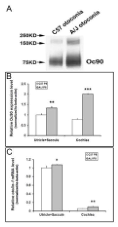
Figure 10.

Otoconial deposition and epithelial expression of otoconial proteins in A/J vs. C57 inner ears as detected by Western blotting (A) and real-time quantitative RT-PCR (B, C). In (A), total otoconia extract from one C57 and one A/J mouse at age P4 was loaded in each lane (labeled as “O”). This increase of Oc90 in A/J otoconia is confirmed by loading an equal amount of total protein in each lane (not shown). (B, C) Real-time PCR shows a significant increase in both Oc90 and Otol1 mRNA in the A/J inner ear epithelia as compared to C57 tissues at age P8 (*, P < 0.05; **, P < 0.01; ***, P < 0.001, n=3).
