Fig. 3.
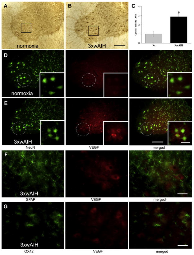
Representative images of VEGF immunostaining in C4 ventral horn. VEGF was expressed in large, NeuN positive cells, including large, presumptive phrenic motor neurons (in black boxes) and interneurons. 3×wAIH significantly increased VEGF protein expression in presumptive phrenic motor neurons versus control, normoxic rats (A–B). Boxes indicate the region in which densitometry was performed; higher-magnification images from this region are in the bottom right corner. Optical density (OD) analysis confirmed significant increase in VEGF immunoreactivity following 3×wAIH (C). Data are means±1 SEM. *p<0.05 versus normoxic controls. Immunofluorescence of large, NeuN positive cells (green) confirmed that VEGF (red) is localized in presumptive phrenic motor neurons (in white circle). VEGF immunoreactivity is enhanced following 3×wAIH (D–E). VEGF was not detectable in GFAP positive astrocytes (F) or in OX-42 positive microglia (G), even after 3×wAIH. Scale bars: 200 μm for lower magnification (A–B, D–E) and 100 μm for higher magnification (small boxes in D–E); F–G is 50 μm.

