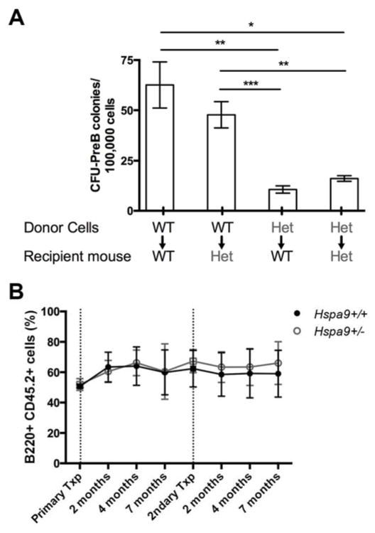
Figure 4. The reduction in Hspa9+/− CFU-PreB colony formation is hematopoietic cell-intrinsic.
A) Donor bone marrow from Hspa9+/+ (WT) or Hspa9+/− (HET) mice was transplanted into lethally irradiated Hspa9+/+ (WT) or Hspa9+/− (HET) recipients. Bone marrow was harvested 6 months after transplant and plated in CFU-PreB promoting methylcellulose (N=4–9 mice/genotype). B) A ratio of 1:1 Hspa9+/+ (black lines) or Hspa9+/− (grey lines) test cells (Ly5.2) and competitor bone marrow (Ly5.1/5.2) were transplanted into lethally irradiated recipients (Ly5.1). Mice were bled at intervals indicated after transplant and relative chimerism of B220+ peripheral blood cells were evaluated in recipients. Following long-term engraftment, bone marrow from recipients were pooled and transplanted into lethally irradiated secondary recipients. Data represents pooled results from two independently transplanted cohorts (N=10–15 mice/genotype). Txp, transplant. Statistical analysis by two tailed Student’s t-test and an ANOVA. Error bars represent mean ± SD. *p<0.05, **p<0.01, ***p<0.001

