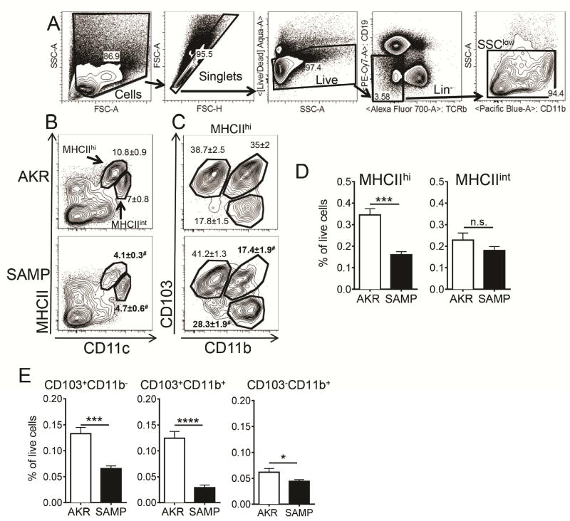
Figure 1. Defective MLN DC in SAMP mice.
(A) Single cell suspensions were prepared by incubating minced tissues in collagenase and DNAse I. Sequential gates were set to exclude debris, doublets, dead cells, cells of B and T cell lineage and granulocytes (SSChi). (B) Representative flow cytometry analysis of DC in AKR and SAMP MLN from 10–20 weeks old mice. Two populations of CD11c+MHCIIhi and CD11chiMHCIIint MLN DCs were detected. (C) MHCIIhi cells contained 3 major subpopulations: CD103+CD11b+, CD103+CD11b− and CD103−CD11b− cells. Mean of percentages of parent gate ± SEM are indicated on the plots, # denotes statistically significant differences between AKR and SAMP mice. (D) The fraction of MHCIIhi and MHCIIint cells and (E) subpopulations of MHCIIhiCD11c+ cells was calculated as % of all live cells. Results are the mean ± SEM of 3 experiments with n=5 in AKR and n=6 in SAMP groups. n.s – not significant, **** p<0.0001, *** p<0.001, ** p<0.01, * p<0.05 by two-sided Student’s t test

