Fig. 2.
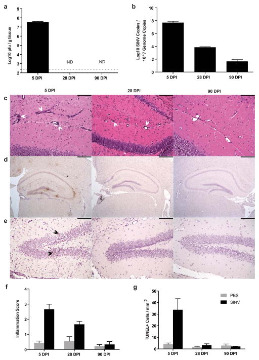
SINV, inflammation, and cell death in the brain over time. (a) Infectious SINV was detectable by plaque assay at high levels in the brain at 5 DPI but undetectable at 28 and 90 DPI (dotted line represents limit of detection; ND = not detectable). (b) SINV RNA levels were highest at 5 DPI, lower at 28 DPI, and just above the level of detection at 90 DPI in the brain. (c) Representative photomicrographs of H&E-stained coronal brain sections from SINV-infected mice demonstrating the levels of parenchymal inflammation and perivascular cuffing in the hippocampus (white arrowheads denote perivascular cuffing; 200X magnification; scale bar = 100μm). (d) Representative photomicrographs of immunohistochemical staining for SINV antigen in infected mice at each time point. SINV protein was readily detectable in the hippocampus at 5 DPI, but not at 28 or 90 DPI (brown staining = SINV protein; 40X magnification; scale bar = 500μm). (e) Representative photomicrographs from TUNEL staining of the hippocampal dentate gyrus of SINV-infected mice at each time point. Multiple apoptotic cells were detected at 5 DPI but not at 28 and 90 DPI (brown nuclear staining = TUNEL-positive [denoted by black arrowheads]; 200X magnification; scale bar = 100μm). (f) Quantification of brain inflammation. Inflammation was greater at 5 and 28 DPI in SINV-infected than mock-infected control mice. (g) Quantification of TUNEL-positive cells in the hippocampus. Higher numbers of apoptotic/necrotic cells were detected in SINV-infected mice compared to mock-infected control mice at 5 DPI, but not at 28 or 90 DPI (For all bar graphs, N=3–4 mice per group per time point; data presented as mean ± SEM)

