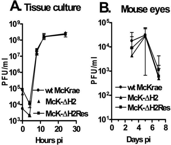
Fig. 7.
Replication of McK-ΔH2 in tissue culture and in rabbit eyes. Panel A: RS cell monolayers were infected with the indicated virus at an MOI of 0.01. At the times indicated the monolayer and tissue culture media were freeze thawed 2X and the amount of virus determined by standard plaque assays on RS cells. Each time point was done in triplicate. Panel B: Rabbits were ocularly infected with 2 × 10⁵ pfu/eye of the indicated virus. Tears swabs were collected on the indicated days and the amount of virus determined by plaque assays on RS cells. Each time point represents the average of 10 eyes.

