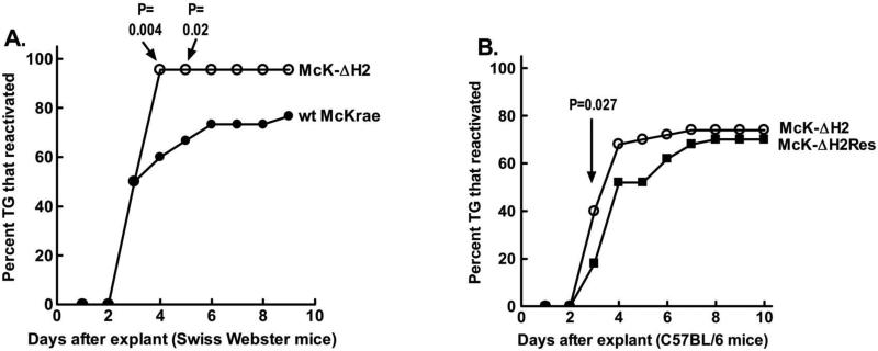
Fig. 9.
Reactivation of McK-ΔH2. Panel A: Swiss Webster mice were ocularly infected with 2×10⁵ pfu/eye of the indicated virus. 30 days pi surviving mice were euthanized and TG removed for induction of reactivation by explantation into tissue culture media. Aliquots of the tissue culture media were collected daily and plated on RS indicator cells to determine the time of first appearance of reactivated virus. The cumulative percent of TG from which virus had reactivated are plotted. Wt McKrae: 30 TG; McK-ΔH2: 22 TG. Panel B: C57BL/6 mice were infected with the indicated virus except that 1×10⁶ pfu/eye were used, and reactivation determined as in panel A. 50 TG per group.

