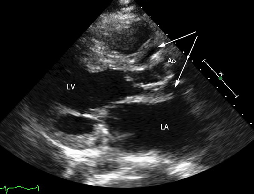
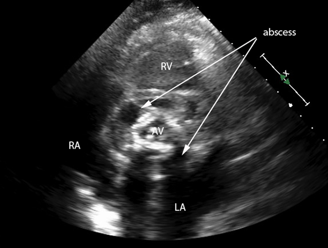
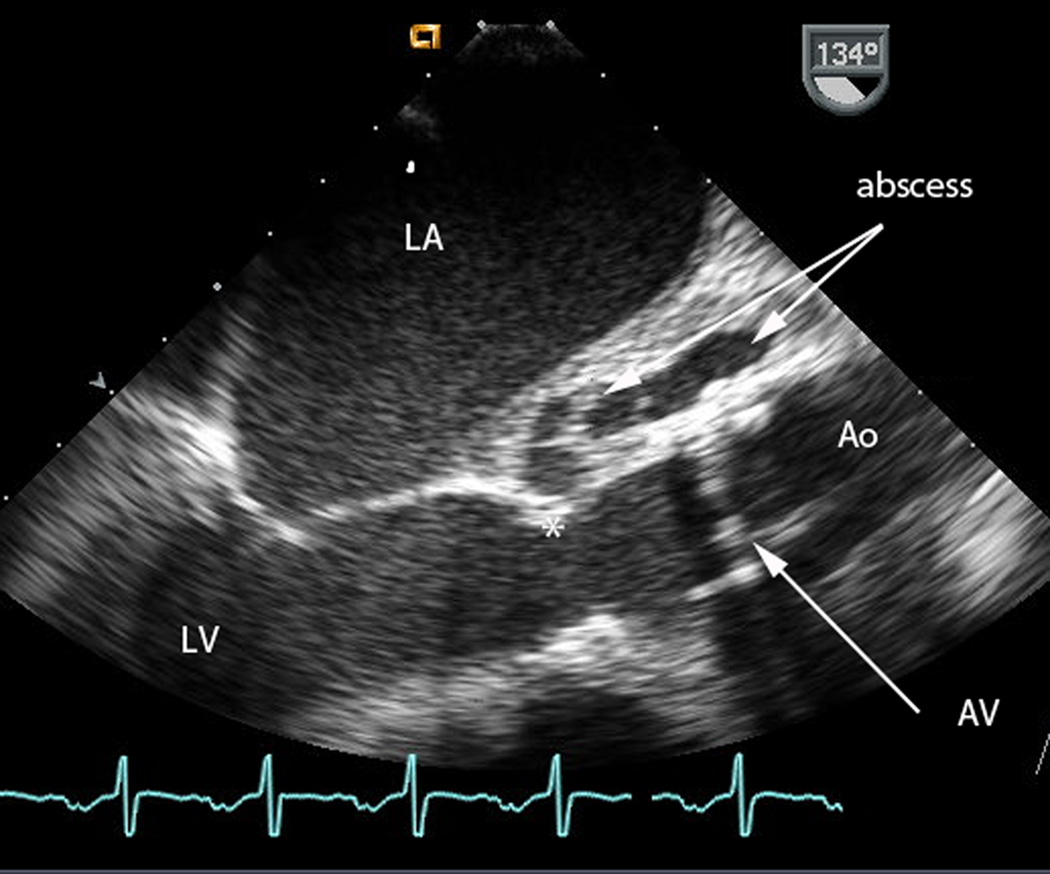
Figure 1.
a) Transthoracic echocardiogram, parasternal long axis view. The left ventricle (LV) ejects to the ascending aorta (Ao). Surrounding the aortic root is a large abscess (arrows).
b) Transthoracic echocardiogram, parasternal short axis view. Measurement markers along the right of the image represent 1 cm distances. The aortic valve (AV) is seen in the center of the image. Surrounding the valve is a large, complex abscess (arrows) which extends 1–2 cm circumferentially around the AV.
c) Intraoperative transesophageal echocardiogram, taken just before placement of the total artificial heart. Measurement markers along the right of the image represent 1 cm distances. The mechanical aortic valve (AV) is dehisced from the aortic valve attachment (*), and a large abscess (arrows) is seen in the aortomitral intervalvular fibrosa.


