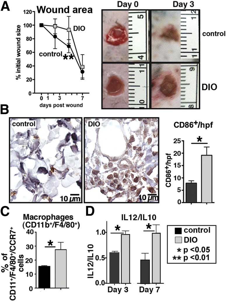
Figure 2.
Delayed wound healing in DIO mice is associated with increased proinflammatory macrophages. Punch biopsies (4 mm) were performed on the back of DIO and control mice. Change in wound area was recorded daily using ImageJ software (National Institutes of Health) until complete healing was observed. A: Wound healing curves in DIO and control mice (n = 10/time point). Data are pooled from three experiments and are expressed as mean ± SD. Representative images of wounds at day 0 and 3 days postwounding are shown. B: Immunohistochemical analysis of mononuclear cells (CD86+) in wounds at day 3 (cells/high-powered field [hpf]) in DIO compared with controls (n = 6). Representative examples of DIO and control wounds are shown. C: Flow cytometry of DIO and control wounds at day 3. Proinflammatory macrophages were defined as CD11b+/F4/80+ cells that coexpressed CCR7 (n = 6, experiment replicated once). D: Ratio of IL-12 (M1) to IL-10 (M2) cytokine levels in wounds at days 3 and 7 analyzed by Bioplex (n = 4, experiment replicated one time). Data are expressed as mean ± SE.

