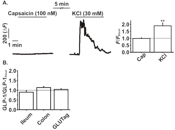
Figure 7. Analysis of functional TRPV1 in primary enteroendocrine cells and GLUTag cells.

A. Addition of capsaicin (Cap, 100 nM) failed to elicit any significant change in intracellular Ca2+ levels as measured by GCaMP3 fluorescence in colonic L-cells from GLU-Cre/ROSA26-GCaMP3 mice. KCl (30 mM) was used to confirm the responsiveness of each L-cell analysed. B. Incubation of murine ileal or colonic intestinal cultures, or GLUTag cells, with capsaicin (100 nM) for two hours did not significantly alter GLP-1 secretion compared to basal (10 mM glucose). All experiments were n= ≥5, *p<0.05, repeated measures ANOVA or one-way ANOVA, both with Bonferroni post-hoc test. The dotted line on each graph represents the respective baseline value.
