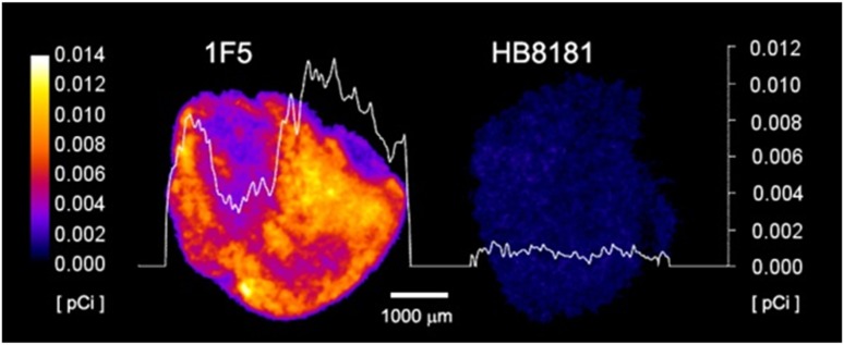
Figure 3.
α-Camera imaging of subcutaneous Ramos xenografts. Images obtained 28 hours after IV injection of [211At]1F5-B10 (anti-CD20, left) or [211At]HB8181-B10 (control, right), demonstrating specificity of CD20 targeting but heterogeneous dose distributions. Images are color coded to express the intratumoral activity in pCi per voxel (17 × 17 × 16 μm). The white curve represents the activity variation along a line profile placed centrally in each tumor. The white bar (bottom center) indicates 1000 μm.

