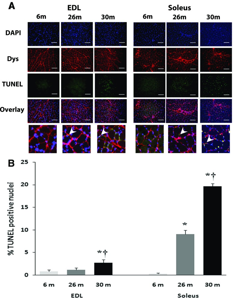
Fig. 4.
Quantification of apoptosis with age is shown in soleus and EDL of female F344BN rats. a Representative images of the triple staining (DAPI, Dystrophin, TUNEL, and an overlay of the three) for EDL (left) and soleus (right) muscle sections from 6-, 26-, and 30-month female FBN rats. Apoptotic myonuclei were visualized with TUNEL staining. Muscle borders were visualized using mouse monoclonal antibody dystrophin (C-terminus), and all nuclei were stained with 4′, 6-diamidino-2-phenyllindole (DAPI). Arrows in magnified overlays indicate TUNEL-positive nuclei. b Graph representing the TUNEL-positive nuclei in EDL and soleus muscle sections. Data are presented as mean ± SEM. *P < 0.05 indicates significant difference from young adult (6-month) age group. † P < 0.05 indicates significant difference from aged (26-month) group. n = 3 each group

