FIG 6.

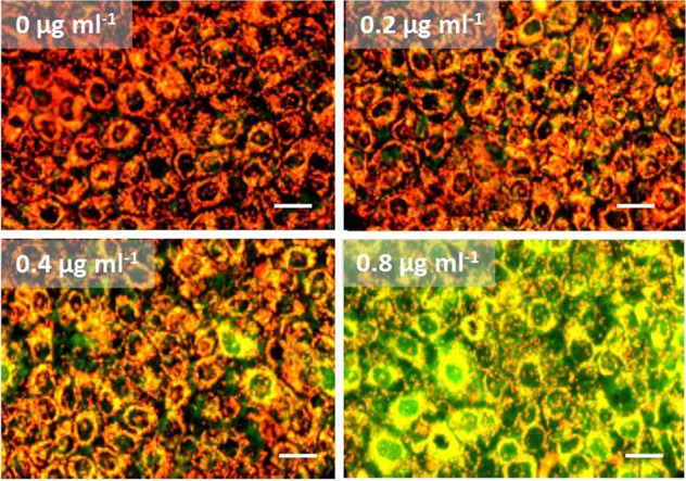
Purified amylosin causes a dose-dependent loss in the cellular transmembrane potential (ΔΨ) in human keratinocytes (HaCaT). Monolayers of HaCaT cells were exposed to 0, 0.2, 0.4, or 0.8 μg of purified amylosin ml−1 for 24 h and then double-stained with the membrane-permeant ΔΨ-responsive dye JC-1 and the membrane-nonpermeant dye propidium iodide. The orange fluorescence in JC-1-stained cells indicates a high membrane potential (ΔΨ, >140 mV), and green fluorescence indicates a dissipated membrane potential (ΔΨ, <100 mV). The 0 μg ml−1 data are for the vehicle-only control (≤1% [vol/vol] methanol). None of the cells displayed purple-red fluorescence following propidium iodide staining (death staining). The images are representative of three independent microscopic views. Bar, 30 μm.
