FIG 4.

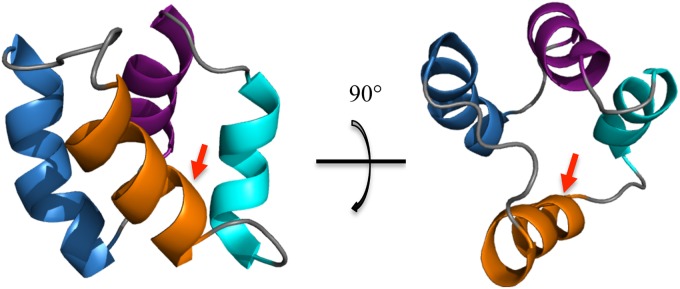
NMR solution structure of acidocin B (PDB code 2MWR): helix 1 is in blue, helix 2 is in purple, helix 3 is in cyan, and helix 4 is in orange. The arrow indicates the linkage of the N and C termini.

NMR solution structure of acidocin B (PDB code 2MWR): helix 1 is in blue, helix 2 is in purple, helix 3 is in cyan, and helix 4 is in orange. The arrow indicates the linkage of the N and C termini.