Figure 2.
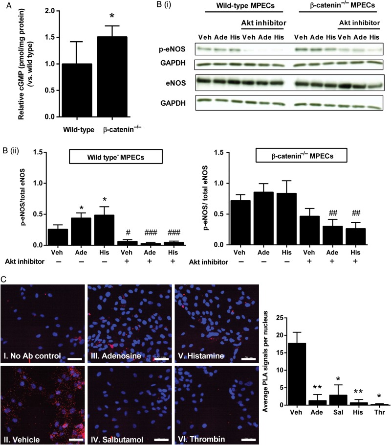
Loss of β-catenin increases basal eNOS phosphorylation and cGMP production. (A) cGMP production was assessed in wild-type and β-catenin−/− MPECs by ELISA and results expressed relative to protein content per sample (shown relative to wild-type; *P < 0.05, unpaired t-test, n = 4 per group). (B) Wild-type and β-catenin−/− MPECs were treated with adenosine (100 μmol/L), histamine (100 μmol/L), or vehicle for 2.5 min in the presence or absence of Akt inhibitor (10 μmol/L; pre-treatment for 30 min). B(i), Cell lysates were probed by western blotting for phosphoSer-1177-eNOS and total eNOS, and B(ii) results expressed as the densitometric ratio of phosphoSer-1177-eNOS/GAPDH to total eNOS/GAPDH (*P < 0.05 vs. vehicle, #P < 0.05, ###P < 0.001 vs. corresponding treatment in the absence of Akt inhibitor; two-way ANOVA; n = 6 per group). Wild-type vehicle vs. β-catenin−/− vehicle were analysed separately by unpaired t-test (**P < 0.01). (C) HUVECs were treated with adenosine (100 μmol/L), salbutamol (1 µmol/L), histamine (100 μmol/L), thrombin (1 IU/mL), or vehicle for 2.5 min then fixed and subject to PLA using antibodies targeting eNOS and β-catenin (II–IV). PLA was also carried out with the omission of primary antibodies (I). Images are representative of three independent cultures (scale bar shows 50 µm). Images were quantified using ImageJ Particle Analyser to determine the number of PLA signal per image and expressed as the mean number of signals per nucleus (*P < 0.05, **P < 0.01, one-way ANOVA with repeated measures, n = 3 per group).

