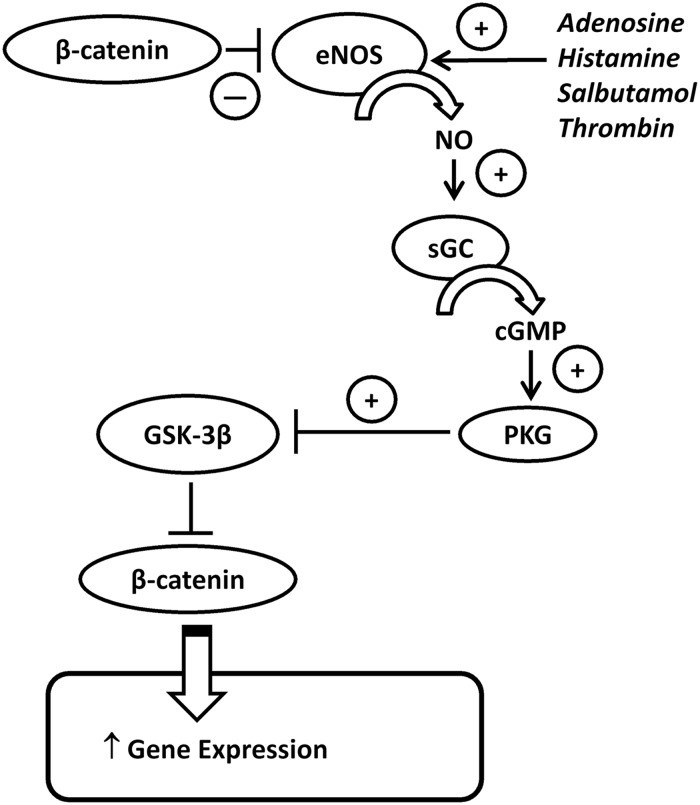
Figure 7.
Schematic diagram of NO-β-catenin cross-regulation. In unstimulated cells, β-catenin is excluded from the nucleus due to the inhibitory action of GSK-3β (which phosphorylates β-catenin targeting it for degradation), while membrane-associated β-catenin binds to and inhibits eNOS activity. Following activation of eNOS using agonists, i.e. adenosine, histamine, salbutamol or thrombin, this association with β-catenin is lost. At the same time, agonists increase eNOS activity and NO production which increases the activity of soluble guanylate cyclase leading to increased production of cGMP which consequently increases the activity of PKG. PKG phosphorylates and inhibits GSK-3β which permits activation of β-catenin. Active (dephosphorylated) β-catenin is able to translocate to the nucleus where it exerts effects on gene transcription. The effects of eNOS activation are indicated by plus or minus symbols.

